Iron »
PDB 1sdl-1stq »
1smi »
Iron in PDB 1smi: A Single Mutation of P450 BM3 Induces the Conformational Rearrangement Seen Upon Substrate-Binding in Wild-Type Enzyme
Enzymatic activity of A Single Mutation of P450 BM3 Induces the Conformational Rearrangement Seen Upon Substrate-Binding in Wild-Type Enzyme
All present enzymatic activity of A Single Mutation of P450 BM3 Induces the Conformational Rearrangement Seen Upon Substrate-Binding in Wild-Type Enzyme:
1.14.14.1;
1.14.14.1;
Protein crystallography data
The structure of A Single Mutation of P450 BM3 Induces the Conformational Rearrangement Seen Upon Substrate-Binding in Wild-Type Enzyme, PDB code: 1smi
was solved by
M.G.Joyce,
H.M.Girvan,
A.W.Munro,
D.Leys,
with X-Ray Crystallography technique. A brief refinement statistics is given in the table below:
| Resolution Low / High (Å) | 14.92 / 2.00 |
| Space group | P 21 21 21 |
| Cell size a, b, c (Å), α, β, γ (°) | 61.206, 118.929, 146.338, 90.00, 90.00, 90.00 |
| R / Rfree (%) | 22.4 / 28.8 |
Iron Binding Sites:
The binding sites of Iron atom in the A Single Mutation of P450 BM3 Induces the Conformational Rearrangement Seen Upon Substrate-Binding in Wild-Type Enzyme
(pdb code 1smi). This binding sites where shown within
5.0 Angstroms radius around Iron atom.
In total 2 binding sites of Iron where determined in the A Single Mutation of P450 BM3 Induces the Conformational Rearrangement Seen Upon Substrate-Binding in Wild-Type Enzyme, PDB code: 1smi:
Jump to Iron binding site number: 1; 2;
In total 2 binding sites of Iron where determined in the A Single Mutation of P450 BM3 Induces the Conformational Rearrangement Seen Upon Substrate-Binding in Wild-Type Enzyme, PDB code: 1smi:
Jump to Iron binding site number: 1; 2;
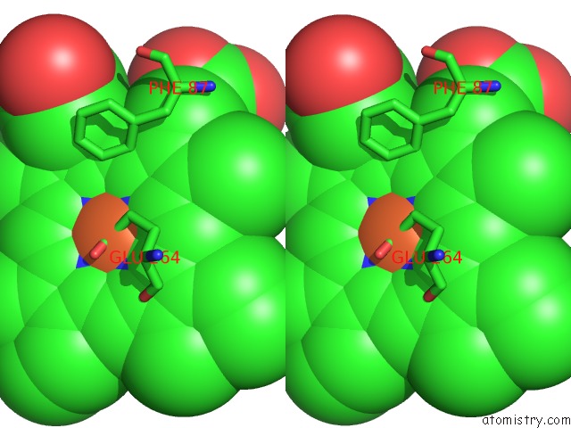
Iron binding site 1 out of 2 in 1smi
Go back to
Iron binding site 1 out
of 2 in the A Single Mutation of P450 BM3 Induces the Conformational Rearrangement Seen Upon Substrate-Binding in Wild-Type Enzyme
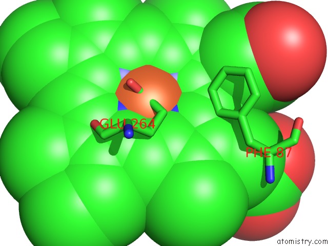
Mono view
Stereo pair view
Mono view
Stereo pair view
A full contact list of Iron with other atoms in the Fe binding
site number 1 of A Single Mutation of P450 BM3 Induces the Conformational Rearrangement Seen Upon Substrate-Binding in Wild-Type Enzyme within 5.0Å range:
|
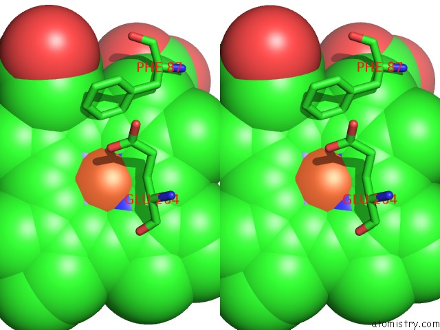
Iron binding site 2 out of 2 in 1smi
Go back to
Iron binding site 2 out
of 2 in the A Single Mutation of P450 BM3 Induces the Conformational Rearrangement Seen Upon Substrate-Binding in Wild-Type Enzyme
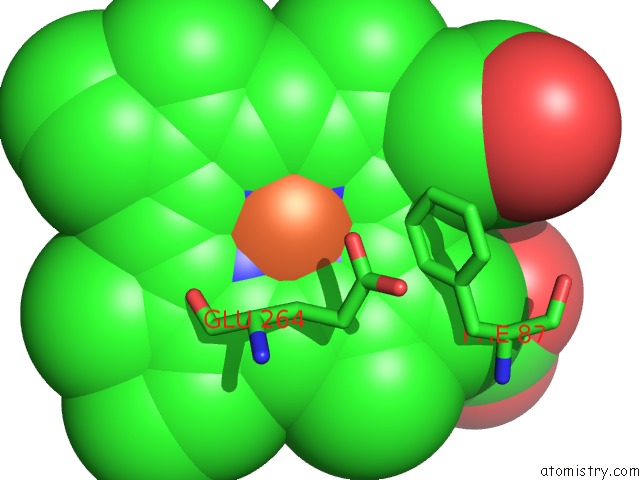
Mono view
Stereo pair view
Mono view
Stereo pair view
A full contact list of Iron with other atoms in the Fe binding
site number 2 of A Single Mutation of P450 BM3 Induces the Conformational Rearrangement Seen Upon Substrate-Binding in Wild-Type Enzyme within 5.0Å range:
|
Reference:
M.G.Joyce,
H.M.Girvan,
A.W.Munro,
D.Leys.
A Single Mutation in Cytochrome P450 BM3 Induces the Conformational Rearrangement Seen Upon Substrate Binding in the Wild-Type Enzyme J.Biol.Chem. V. 279 23287 2004.
ISSN: ISSN 0021-9258
PubMed: 15020590
DOI: 10.1074/JBC.M401717200
Page generated: Wed Jul 16 20:41:52 2025
ISSN: ISSN 0021-9258
PubMed: 15020590
DOI: 10.1074/JBC.M401717200
Last articles
Fe in 2YXOFe in 2YRS
Fe in 2YXC
Fe in 2YNM
Fe in 2YVJ
Fe in 2YP1
Fe in 2YU2
Fe in 2YU1
Fe in 2YQB
Fe in 2YOO