Iron »
PDB 1sdl-1stq »
1sof »
Iron in PDB 1sof: Crystal Structure of the Azotobacter Vinelandii Bacterioferritin at 2.6 A Resolution
Protein crystallography data
The structure of Crystal Structure of the Azotobacter Vinelandii Bacterioferritin at 2.6 A Resolution, PDB code: 1sof
was solved by
H.L.Liu,
J.F.Huang,
R.C.Bi,
with X-Ray Crystallography technique. A brief refinement statistics is given in the table below:
| Resolution Low / High (Å) | 25.32 / 2.60 |
| Space group | H 3 |
| Cell size a, b, c (Å), α, β, γ (°) | 124.965, 124.965, 287.406, 90.00, 90.00, 120.00 |
| R / Rfree (%) | 19.6 / 24.6 |
Other elements in 1sof:
The structure of Crystal Structure of the Azotobacter Vinelandii Bacterioferritin at 2.6 A Resolution also contains other interesting chemical elements:
| Magnesium | (Mg) | 10 atoms |
| Barium | (Ba) | 2 atoms |
Iron Binding Sites:
Pages:
>>> Page 1 <<< Page 2, Binding sites: 11 - 20;Binding sites:
The binding sites of Iron atom in the Crystal Structure of the Azotobacter Vinelandii Bacterioferritin at 2.6 A Resolution (pdb code 1sof). This binding sites where shown within 5.0 Angstroms radius around Iron atom.In total 20 binding sites of Iron where determined in the Crystal Structure of the Azotobacter Vinelandii Bacterioferritin at 2.6 A Resolution, PDB code: 1sof:
Jump to Iron binding site number: 1; 2; 3; 4; 5; 6; 7; 8; 9; 10;
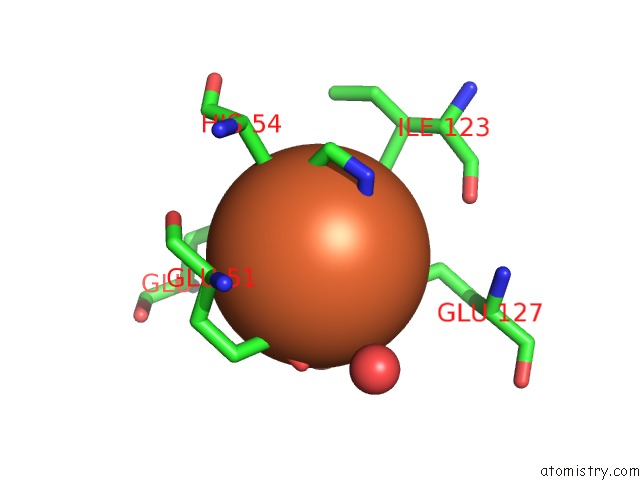
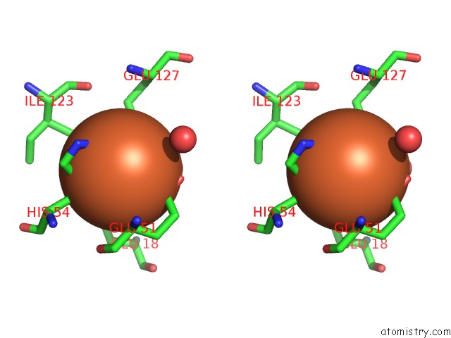
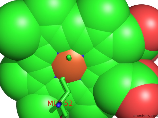
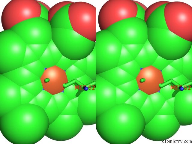
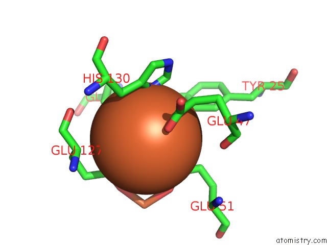
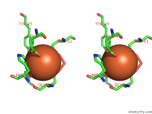
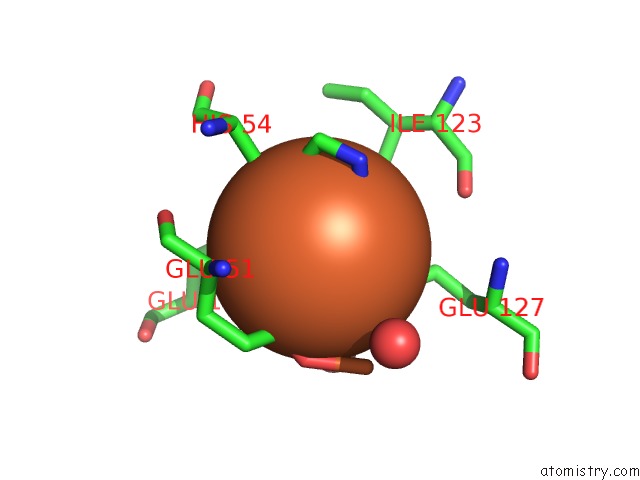
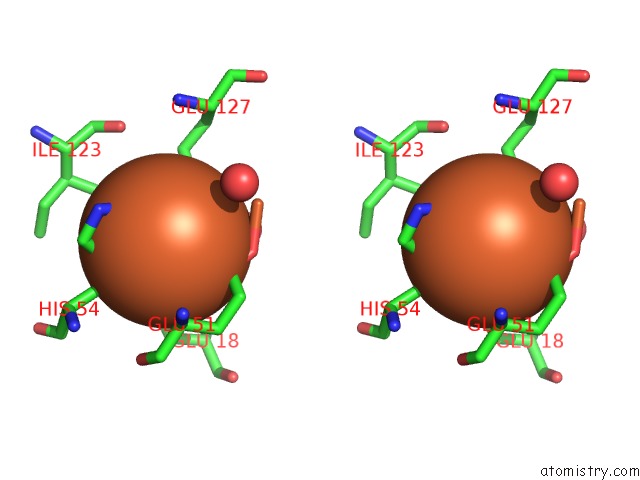
Iron binding site 1 out of 20 in 1sof
Go back to
Iron binding site 1 out
of 20 in the Crystal Structure of the Azotobacter Vinelandii Bacterioferritin at 2.6 A Resolution
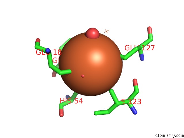
Mono view
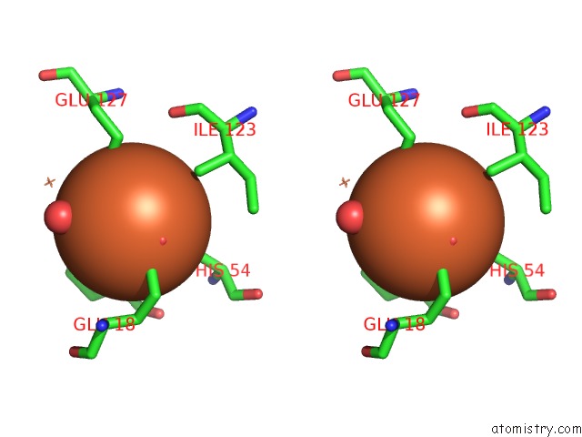
Stereo pair view
Mono view
Stereo pair view
|
|
A full contact list of Iron with other atoms in the Fe binding
site number 1 of Crystal Structure of the Azotobacter Vinelandii Bacterioferritin at 2.6 A Resolution within 5.0Å range:
|
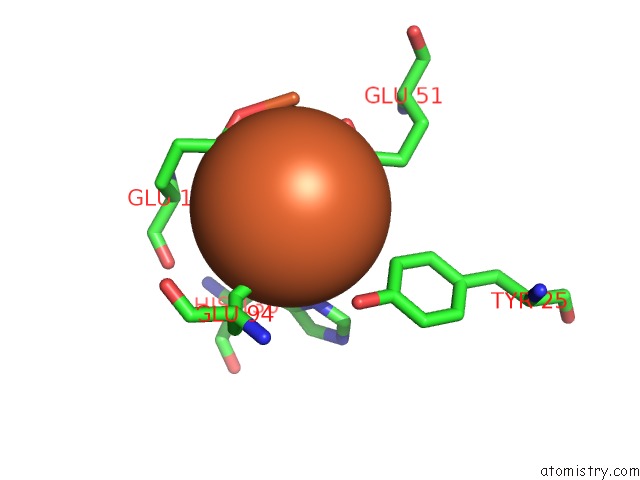
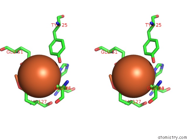
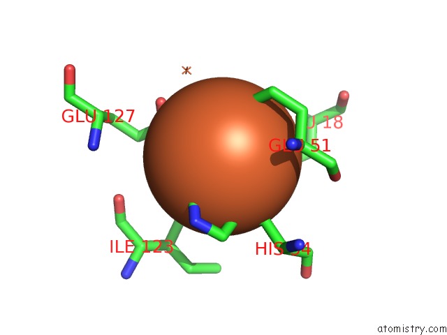
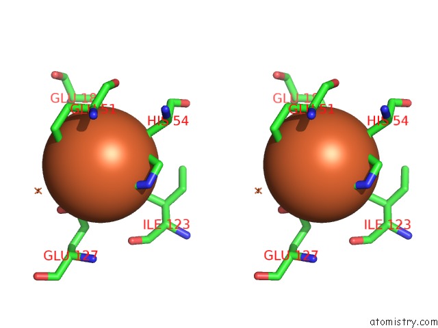
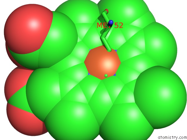
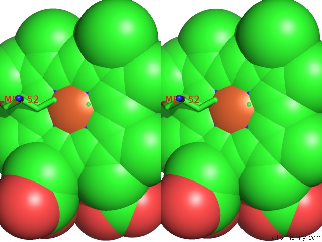
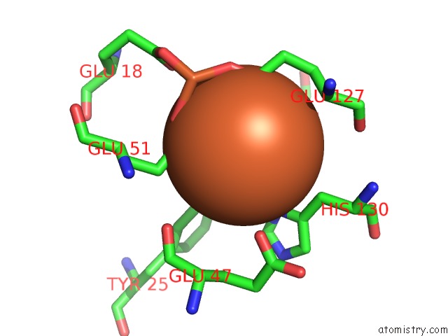
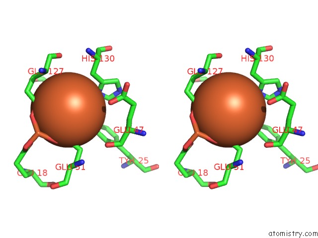
Iron binding site 2 out of 20 in 1sof
Go back to
Iron binding site 2 out
of 20 in the Crystal Structure of the Azotobacter Vinelandii Bacterioferritin at 2.6 A Resolution
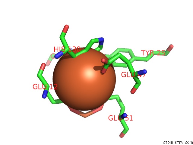
Mono view
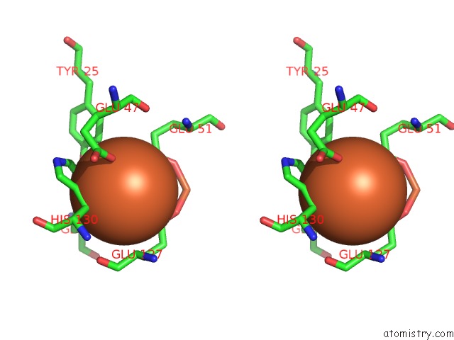
Stereo pair view
Mono view
Stereo pair view
|
|
A full contact list of Iron with other atoms in the Fe binding
site number 2 of Crystal Structure of the Azotobacter Vinelandii Bacterioferritin at 2.6 A Resolution within 5.0Å range:
|
Iron binding site 3 out of 20 in 1sof
Go back to
Iron binding site 3 out
of 20 in the Crystal Structure of the Azotobacter Vinelandii Bacterioferritin at 2.6 A Resolution
Mono view
Stereo pair view
Mono view
Stereo pair view
|
|
A full contact list of Iron with other atoms in the Fe binding
site number 3 of Crystal Structure of the Azotobacter Vinelandii Bacterioferritin at 2.6 A Resolution within 5.0Å range:
|
Iron binding site 4 out of 20 in 1sof
Go back to
Iron binding site 4 out
of 20 in the Crystal Structure of the Azotobacter Vinelandii Bacterioferritin at 2.6 A Resolution
Mono view
Stereo pair view
Mono view
Stereo pair view
|
|
A full contact list of Iron with other atoms in the Fe binding
site number 4 of Crystal Structure of the Azotobacter Vinelandii Bacterioferritin at 2.6 A Resolution within 5.0Å range:
|
Iron binding site 5 out of 20 in 1sof
Go back to
Iron binding site 5 out
of 20 in the Crystal Structure of the Azotobacter Vinelandii Bacterioferritin at 2.6 A Resolution
Mono view
Stereo pair view
Mono view
Stereo pair view
|
|
A full contact list of Iron with other atoms in the Fe binding
site number 5 of Crystal Structure of the Azotobacter Vinelandii Bacterioferritin at 2.6 A Resolution within 5.0Å range:
|
Iron binding site 6 out of 20 in 1sof
Go back to
Iron binding site 6 out
of 20 in the Crystal Structure of the Azotobacter Vinelandii Bacterioferritin at 2.6 A Resolution
Mono view
Stereo pair view
Mono view
Stereo pair view
|
|
A full contact list of Iron with other atoms in the Fe binding
site number 6 of Crystal Structure of the Azotobacter Vinelandii Bacterioferritin at 2.6 A Resolution within 5.0Å range:
|
Iron binding site 7 out of 20 in 1sof
Go back to
Iron binding site 7 out
of 20 in the Crystal Structure of the Azotobacter Vinelandii Bacterioferritin at 2.6 A Resolution
Mono view
Stereo pair view
Mono view
Stereo pair view
|
|
A full contact list of Iron with other atoms in the Fe binding
site number 7 of Crystal Structure of the Azotobacter Vinelandii Bacterioferritin at 2.6 A Resolution within 5.0Å range:
|
Iron binding site 8 out of 20 in 1sof
Go back to
Iron binding site 8 out
of 20 in the Crystal Structure of the Azotobacter Vinelandii Bacterioferritin at 2.6 A Resolution
Mono view
Stereo pair view
Mono view
Stereo pair view
|
|
A full contact list of Iron with other atoms in the Fe binding
site number 8 of Crystal Structure of the Azotobacter Vinelandii Bacterioferritin at 2.6 A Resolution within 5.0Å range:
|
Iron binding site 9 out of 20 in 1sof
Go back to
Iron binding site 9 out
of 20 in the Crystal Structure of the Azotobacter Vinelandii Bacterioferritin at 2.6 A Resolution
Mono view
Stereo pair view
Mono view
Stereo pair view
|
|
A full contact list of Iron with other atoms in the Fe binding
site number 9 of Crystal Structure of the Azotobacter Vinelandii Bacterioferritin at 2.6 A Resolution within 5.0Å range:
|
Iron binding site 10 out of 20 in 1sof
Go back to
Iron binding site 10 out
of 20 in the Crystal Structure of the Azotobacter Vinelandii Bacterioferritin at 2.6 A Resolution
Mono view
Stereo pair view
Mono view
Stereo pair view
|
|
A full contact list of Iron with other atoms in the Fe binding
site number 10 of Crystal Structure of the Azotobacter Vinelandii Bacterioferritin at 2.6 A Resolution within 5.0Å range:
|
Reference:
H.L.Liu,
H.N.Zhou,
W.M.Xing,
J.F.Zhao,
S.X.Li,
J.F.Huang,
R.C.Bi.
2.6 A Resolution Crystal Structure of the Bacterioferritin From Azotobacter Vinelandii Febs Lett. V. 573 93 2004.
ISSN: ISSN 0014-5793
PubMed: 15327981
DOI: 10.1016/J.FEBSLET.2004.07.054
Page generated: Wed Jul 16 20:43:03 2025
ISSN: ISSN 0014-5793
PubMed: 15327981
DOI: 10.1016/J.FEBSLET.2004.07.054
Last articles
Mg in 8S8GMg in 8S8D
Mg in 8S8E
Mg in 8S8F
Mg in 8S1P
Mg in 8RW1
Mg in 8S87
Mg in 8S8B
Mg in 8S8A
Mg in 8S7G