Iron »
PDB 1sdl-1stq »
1spg »
Iron in PDB 1spg: Carbonmonoxy Hemoglobin From the Teleost Fish Leiostomus Xanthurus
Protein crystallography data
The structure of Carbonmonoxy Hemoglobin From the Teleost Fish Leiostomus Xanthurus, PDB code: 1spg
was solved by
S.E.Mylvaganam,
E.D.Getzoff,
with X-Ray Crystallography technique. A brief refinement statistics is given in the table below:
| Resolution Low / High (Å) | 10.00 / 1.95 |
| Space group | C 1 2 1 |
| Cell size a, b, c (Å), α, β, γ (°) | 89.600, 75.600, 69.700, 90.00, 141.90, 90.00 |
| R / Rfree (%) | 19.1 / 24.5 |
Iron Binding Sites:
The binding sites of Iron atom in the Carbonmonoxy Hemoglobin From the Teleost Fish Leiostomus Xanthurus
(pdb code 1spg). This binding sites where shown within
5.0 Angstroms radius around Iron atom.
In total 2 binding sites of Iron where determined in the Carbonmonoxy Hemoglobin From the Teleost Fish Leiostomus Xanthurus, PDB code: 1spg:
Jump to Iron binding site number: 1; 2;
In total 2 binding sites of Iron where determined in the Carbonmonoxy Hemoglobin From the Teleost Fish Leiostomus Xanthurus, PDB code: 1spg:
Jump to Iron binding site number: 1; 2;
Iron binding site 1 out of 2 in 1spg
Go back to
Iron binding site 1 out
of 2 in the Carbonmonoxy Hemoglobin From the Teleost Fish Leiostomus Xanthurus

Mono view
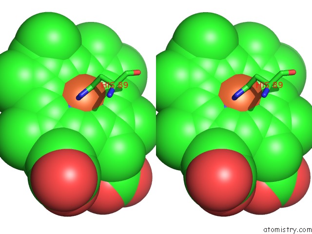
Stereo pair view
Mono view
Stereo pair view
A full contact list of Iron with other atoms in the Fe binding
site number 1 of Carbonmonoxy Hemoglobin From the Teleost Fish Leiostomus Xanthurus within 5.0Å range:
|
Iron binding site 2 out of 2 in 1spg
Go back to
Iron binding site 2 out
of 2 in the Carbonmonoxy Hemoglobin From the Teleost Fish Leiostomus Xanthurus
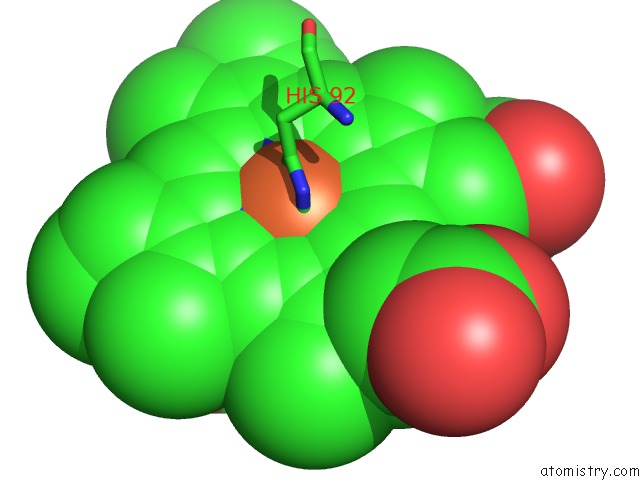
Mono view

Stereo pair view
Mono view
Stereo pair view
A full contact list of Iron with other atoms in the Fe binding
site number 2 of Carbonmonoxy Hemoglobin From the Teleost Fish Leiostomus Xanthurus within 5.0Å range:
|
Reference:
S.E.Mylvaganam,
C.Bonaventura,
J.Bonaventura,
E.D.Getzoff.
Structural Basis For the Root Effect in Haemoglobin. Nat.Struct.Biol. V. 3 275 1996.
ISSN: ISSN 1072-8368
PubMed: 8605630
DOI: 10.1038/NSB0396-275
Page generated: Sat Aug 3 14:48:43 2024
ISSN: ISSN 1072-8368
PubMed: 8605630
DOI: 10.1038/NSB0396-275
Last articles
Zn in 9MJ5Zn in 9HNW
Zn in 9G0L
Zn in 9FNE
Zn in 9DZN
Zn in 9E0I
Zn in 9D32
Zn in 9DAK
Zn in 8ZXC
Zn in 8ZUF