Iron »
PDB 1sdl-1stq »
1sqi »
Iron in PDB 1sqi: Structural Basis For Inhibitor Selectivity Revealed By Crystal Structures of Plant and Mammalian 4- Hydroxyphenylpyruvate Dioxygenases
Protein crystallography data
The structure of Structural Basis For Inhibitor Selectivity Revealed By Crystal Structures of Plant and Mammalian 4- Hydroxyphenylpyruvate Dioxygenases, PDB code: 1sqi
was solved by
C.Yang,
J.W.Pflugrath,
D.L.Camper,
M.L.Foster,
D.J.Pernich,
T.A.Walsh,
with X-Ray Crystallography technique. A brief refinement statistics is given in the table below:
| Resolution Low / High (Å) | 15.00 / 2.15 |
| Space group | P 21 21 21 |
| Cell size a, b, c (Å), α, β, γ (°) | 61.611, 107.481, 133.029, 90.00, 90.00, 90.00 |
| R / Rfree (%) | 21.6 / 27.1 |
Iron Binding Sites:
The binding sites of Iron atom in the Structural Basis For Inhibitor Selectivity Revealed By Crystal Structures of Plant and Mammalian 4- Hydroxyphenylpyruvate Dioxygenases
(pdb code 1sqi). This binding sites where shown within
5.0 Angstroms radius around Iron atom.
In total 4 binding sites of Iron where determined in the Structural Basis For Inhibitor Selectivity Revealed By Crystal Structures of Plant and Mammalian 4- Hydroxyphenylpyruvate Dioxygenases, PDB code: 1sqi:
Jump to Iron binding site number: 1; 2; 3; 4;
In total 4 binding sites of Iron where determined in the Structural Basis For Inhibitor Selectivity Revealed By Crystal Structures of Plant and Mammalian 4- Hydroxyphenylpyruvate Dioxygenases, PDB code: 1sqi:
Jump to Iron binding site number: 1; 2; 3; 4;
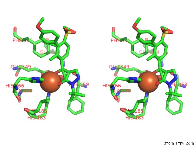
Iron binding site 1 out of 4 in 1sqi
Go back to
Iron binding site 1 out
of 4 in the Structural Basis For Inhibitor Selectivity Revealed By Crystal Structures of Plant and Mammalian 4- Hydroxyphenylpyruvate Dioxygenases

Mono view
Stereo pair view
Mono view
Stereo pair view
A full contact list of Iron with other atoms in the Fe binding
site number 1 of Structural Basis For Inhibitor Selectivity Revealed By Crystal Structures of Plant and Mammalian 4- Hydroxyphenylpyruvate Dioxygenases within 5.0Å range:
|
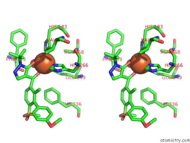
Iron binding site 2 out of 4 in 1sqi
Go back to
Iron binding site 2 out
of 4 in the Structural Basis For Inhibitor Selectivity Revealed By Crystal Structures of Plant and Mammalian 4- Hydroxyphenylpyruvate Dioxygenases

Mono view

Stereo pair view
Mono view
Stereo pair view
A full contact list of Iron with other atoms in the Fe binding
site number 2 of Structural Basis For Inhibitor Selectivity Revealed By Crystal Structures of Plant and Mammalian 4- Hydroxyphenylpyruvate Dioxygenases within 5.0Å range:
|
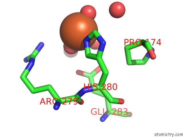
Iron binding site 3 out of 4 in 1sqi
Go back to
Iron binding site 3 out
of 4 in the Structural Basis For Inhibitor Selectivity Revealed By Crystal Structures of Plant and Mammalian 4- Hydroxyphenylpyruvate Dioxygenases

Mono view
Stereo pair view
Mono view
Stereo pair view
A full contact list of Iron with other atoms in the Fe binding
site number 3 of Structural Basis For Inhibitor Selectivity Revealed By Crystal Structures of Plant and Mammalian 4- Hydroxyphenylpyruvate Dioxygenases within 5.0Å range:
|
Iron binding site 4 out of 4 in 1sqi
Go back to
Iron binding site 4 out
of 4 in the Structural Basis For Inhibitor Selectivity Revealed By Crystal Structures of Plant and Mammalian 4- Hydroxyphenylpyruvate Dioxygenases
Mono view

Stereo pair view
Mono view
Stereo pair view
A full contact list of Iron with other atoms in the Fe binding
site number 4 of Structural Basis For Inhibitor Selectivity Revealed By Crystal Structures of Plant and Mammalian 4- Hydroxyphenylpyruvate Dioxygenases within 5.0Å range:
|
Reference:
C.Yang,
J.W.Pflugrath,
D.L.Camper,
M.L.Foster,
D.J.Pernich,
T.A.Walsh.
Structural Basis For Herbicidal Inhibitor Selectivity Revealed By Comparison of Crystal Structures of Plant and Mammalian 4-Hydroxyphenylpyruvate Dioxygenases Biochemistry V. 43 10414 2004.
ISSN: ISSN 0006-2960
PubMed: 15301540
DOI: 10.1021/BI049323O
Page generated: Wed Jul 16 20:47:34 2025
ISSN: ISSN 0006-2960
PubMed: 15301540
DOI: 10.1021/BI049323O
Last articles
Fe in 2YXOFe in 2YRS
Fe in 2YXC
Fe in 2YNM
Fe in 2YVJ
Fe in 2YP1
Fe in 2YU2
Fe in 2YU1
Fe in 2YQB
Fe in 2YOO