Iron »
PDB 1y4r-1yeu »
1y56 »
Iron in PDB 1y56: Crystal Structure of L-Proline Dehydrogenase From P.Horikoshii
Enzymatic activity of Crystal Structure of L-Proline Dehydrogenase From P.Horikoshii
All present enzymatic activity of Crystal Structure of L-Proline Dehydrogenase From P.Horikoshii:
1.5.99.8;
1.5.99.8;
Protein crystallography data
The structure of Crystal Structure of L-Proline Dehydrogenase From P.Horikoshii, PDB code: 1y56
was solved by
H.Tsuge,
R.Kawakami,
H.Sakuraba,
H.Ago,
M.Miyano,
K.Aki,
N.Katunuma,
T.Ohshima,
with X-Ray Crystallography technique. A brief refinement statistics is given in the table below:
| Resolution Low / High (Å) | 19.85 / 2.86 |
| Space group | P 64 2 2 |
| Cell size a, b, c (Å), α, β, γ (°) | 172.219, 172.219, 175.398, 90.00, 90.00, 120.00 |
| R / Rfree (%) | 18.2 / 22.6 |
Other elements in 1y56:
The structure of Crystal Structure of L-Proline Dehydrogenase From P.Horikoshii also contains other interesting chemical elements:
| Chlorine | (Cl) | 1 atom |
Iron Binding Sites:
The binding sites of Iron atom in the Crystal Structure of L-Proline Dehydrogenase From P.Horikoshii
(pdb code 1y56). This binding sites where shown within
5.0 Angstroms radius around Iron atom.
In total only one binding site of Iron was determined in the Crystal Structure of L-Proline Dehydrogenase From P.Horikoshii, PDB code: 1y56:
In total only one binding site of Iron was determined in the Crystal Structure of L-Proline Dehydrogenase From P.Horikoshii, PDB code: 1y56:
Iron binding site 1 out of 1 in 1y56
Go back to
Iron binding site 1 out
of 1 in the Crystal Structure of L-Proline Dehydrogenase From P.Horikoshii
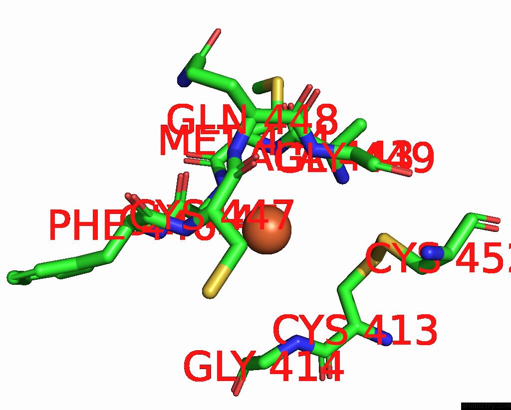
Mono view
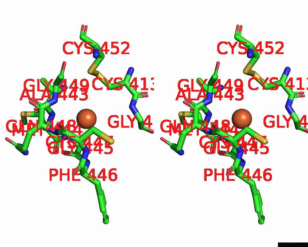
Stereo pair view
Mono view
Stereo pair view
A full contact list of Iron with other atoms in the Fe binding
site number 1 of Crystal Structure of L-Proline Dehydrogenase From P.Horikoshii within 5.0Å range:
|
Reference:
H.Tsuge,
R.Kawakami,
H.Sakuraba,
H.Ago,
M.Miyano,
K.Aki,
N.Katunuma,
T.Ohshima.
Crystal Structure of A Novel Fad-, Fmn-, and Atp-Containing L-Proline Dehydrogenase Complex From Pyrococcus Horikoshii J.Biol.Chem. V. 280 31045 2005.
ISSN: ISSN 0021-9258
PubMed: 16027125
DOI: 10.1074/JBC.C500234200
Page generated: Sat Aug 3 17:18:51 2024
ISSN: ISSN 0021-9258
PubMed: 16027125
DOI: 10.1074/JBC.C500234200
Last articles
Zn in 9J0NZn in 9J0O
Zn in 9J0P
Zn in 9FJX
Zn in 9EKB
Zn in 9C0F
Zn in 9CAH
Zn in 9CH0
Zn in 9CH3
Zn in 9CH1