Iron »
PDB 1y4r-1yeu »
1ycb »
Iron in PDB 1ycb: Distal Pocket Polarity in Ligand Binding to Myoglobin: Deoxy and Carbonmonoxy Forms of A THREONINE68 (E11) Mutant Investigated By X-Ray Crystallography and Infrared Spectroscopy
Protein crystallography data
The structure of Distal Pocket Polarity in Ligand Binding to Myoglobin: Deoxy and Carbonmonoxy Forms of A THREONINE68 (E11) Mutant Investigated By X-Ray Crystallography and Infrared Spectroscopy, PDB code: 1ycb
was solved by
A.D.Cameron,
S.J.Smerdon,
A.J.Wilkinson,
J.Habash,
J.R.Helliwell,
with X-Ray Crystallography technique. A brief refinement statistics is given in the table below:
| Resolution Low / High (Å) | 8.00 / 2.10 |
| Space group | P 1 21 1 |
| Cell size a, b, c (Å), α, β, γ (°) | 124.600, 42.500, 92.000, 90.00, 92.00, 90.00 |
| R / Rfree (%) | n/a / n/a |
Iron Binding Sites:
The binding sites of Iron atom in the Distal Pocket Polarity in Ligand Binding to Myoglobin: Deoxy and Carbonmonoxy Forms of A THREONINE68 (E11) Mutant Investigated By X-Ray Crystallography and Infrared Spectroscopy
(pdb code 1ycb). This binding sites where shown within
5.0 Angstroms radius around Iron atom.
In total 2 binding sites of Iron where determined in the Distal Pocket Polarity in Ligand Binding to Myoglobin: Deoxy and Carbonmonoxy Forms of A THREONINE68 (E11) Mutant Investigated By X-Ray Crystallography and Infrared Spectroscopy, PDB code: 1ycb:
Jump to Iron binding site number: 1; 2;
In total 2 binding sites of Iron where determined in the Distal Pocket Polarity in Ligand Binding to Myoglobin: Deoxy and Carbonmonoxy Forms of A THREONINE68 (E11) Mutant Investigated By X-Ray Crystallography and Infrared Spectroscopy, PDB code: 1ycb:
Jump to Iron binding site number: 1; 2;
Iron binding site 1 out of 2 in 1ycb
Go back to
Iron binding site 1 out
of 2 in the Distal Pocket Polarity in Ligand Binding to Myoglobin: Deoxy and Carbonmonoxy Forms of A THREONINE68 (E11) Mutant Investigated By X-Ray Crystallography and Infrared Spectroscopy
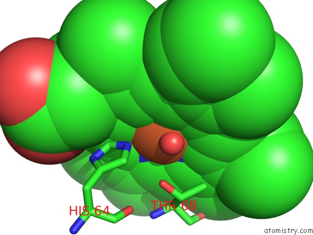
Mono view
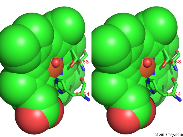
Stereo pair view
Mono view
Stereo pair view
A full contact list of Iron with other atoms in the Fe binding
site number 1 of Distal Pocket Polarity in Ligand Binding to Myoglobin: Deoxy and Carbonmonoxy Forms of A THREONINE68 (E11) Mutant Investigated By X-Ray Crystallography and Infrared Spectroscopy within 5.0Å range:
|
Iron binding site 2 out of 2 in 1ycb
Go back to
Iron binding site 2 out
of 2 in the Distal Pocket Polarity in Ligand Binding to Myoglobin: Deoxy and Carbonmonoxy Forms of A THREONINE68 (E11) Mutant Investigated By X-Ray Crystallography and Infrared Spectroscopy
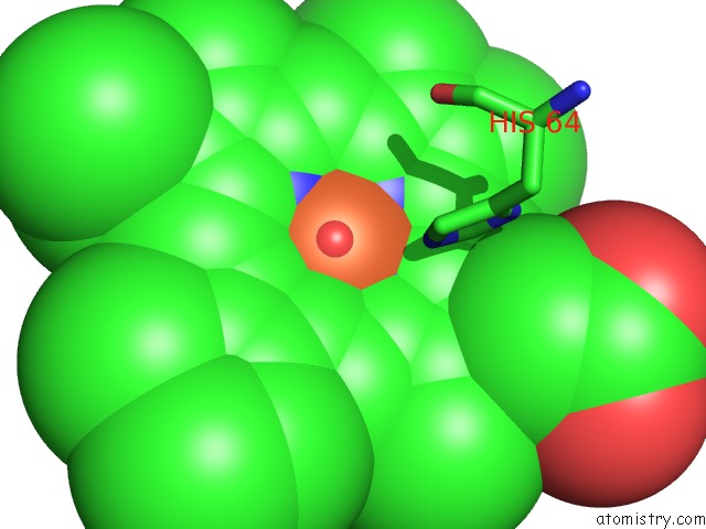
Mono view
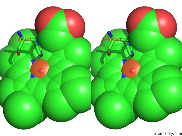
Stereo pair view
Mono view
Stereo pair view
A full contact list of Iron with other atoms in the Fe binding
site number 2 of Distal Pocket Polarity in Ligand Binding to Myoglobin: Deoxy and Carbonmonoxy Forms of A THREONINE68 (E11) Mutant Investigated By X-Ray Crystallography and Infrared Spectroscopy within 5.0Å range:
|
Reference:
A.D.Cameron,
S.J.Smerdon,
A.J.Wilkinson,
J.Habash,
J.R.Helliwell,
T.Li,
J.S.Olson.
Distal Pocket Polarity in Ligand Binding to Myoglobin: Deoxy and Carbonmonoxy Forms of A THREONINE68(E11) Mutant Investigated By X-Ray Crystallography and Infrared Spectroscopy. Biochemistry V. 32 13061 1993.
ISSN: ISSN 0006-2960
PubMed: 8241160
DOI: 10.1021/BI00211A016
Page generated: Sat Aug 3 17:30:04 2024
ISSN: ISSN 0006-2960
PubMed: 8241160
DOI: 10.1021/BI00211A016
Last articles
Zn in 9J0NZn in 9J0O
Zn in 9J0P
Zn in 9FJX
Zn in 9EKB
Zn in 9C0F
Zn in 9CAH
Zn in 9CH0
Zn in 9CH3
Zn in 9CH1