Iron »
PDB 2aiu-2axx »
2amm »
Iron in PDB 2amm: Crystal Structure of L122V/L132V Mutant of Nitrophorin 2
Protein crystallography data
The structure of Crystal Structure of L122V/L132V Mutant of Nitrophorin 2, PDB code: 2amm
was solved by
A.Weichsel,
R.E.Berry,
F.A.Walker,
W.R.Montfort,
with X-Ray Crystallography technique. A brief refinement statistics is given in the table below:
| Resolution Low / High (Å) | 18.00 / 1.90 |
| Space group | P 41 21 2 |
| Cell size a, b, c (Å), α, β, γ (°) | 34.147, 34.147, 256.223, 90.00, 90.00, 90.00 |
| R / Rfree (%) | 14.2 / 22.1 |
Iron Binding Sites:
The binding sites of Iron atom in the Crystal Structure of L122V/L132V Mutant of Nitrophorin 2
(pdb code 2amm). This binding sites where shown within
5.0 Angstroms radius around Iron atom.
In total only one binding site of Iron was determined in the Crystal Structure of L122V/L132V Mutant of Nitrophorin 2, PDB code: 2amm:
In total only one binding site of Iron was determined in the Crystal Structure of L122V/L132V Mutant of Nitrophorin 2, PDB code: 2amm:
Iron binding site 1 out of 1 in 2amm
Go back to
Iron binding site 1 out
of 1 in the Crystal Structure of L122V/L132V Mutant of Nitrophorin 2
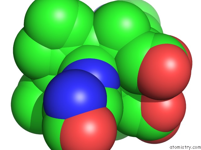
Mono view
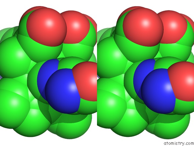
Stereo pair view
Mono view
Stereo pair view
A full contact list of Iron with other atoms in the Fe binding
site number 1 of Crystal Structure of L122V/L132V Mutant of Nitrophorin 2 within 5.0Å range:
|
Reference:
A.Weichsel,
R.E.Berry,
F.A.Walker,
W.R.Montfort.
Crystal Structures, Ligand Induced Conformational Change and Heme Deformation in Complexes of Nitrophorin 2, A Nitric Oxide Transport Protein From Rhodnius Prolixus To Be Published.
Page generated: Sat Aug 3 19:19:39 2024
Last articles
Zn in 9J0NZn in 9J0O
Zn in 9J0P
Zn in 9FJX
Zn in 9EKB
Zn in 9C0F
Zn in 9CAH
Zn in 9CH0
Zn in 9CH3
Zn in 9CH1