Iron »
PDB 2aiu-2axx »
2ani »
Iron in PDB 2ani: Crystal Structure of the F127Y Mutant of Ribonucleotide Reductase R2 From Chlamydia Trachomatis
Enzymatic activity of Crystal Structure of the F127Y Mutant of Ribonucleotide Reductase R2 From Chlamydia Trachomatis
All present enzymatic activity of Crystal Structure of the F127Y Mutant of Ribonucleotide Reductase R2 From Chlamydia Trachomatis:
1.17.4.1;
1.17.4.1;
Protein crystallography data
The structure of Crystal Structure of the F127Y Mutant of Ribonucleotide Reductase R2 From Chlamydia Trachomatis, PDB code: 2ani
was solved by
M.Hogbom,
P.Stenmark,
P.Nordlund,
with X-Ray Crystallography technique. A brief refinement statistics is given in the table below:
| Resolution Low / High (Å) | 20.00 / 2.00 |
| Space group | P 43 21 2 |
| Cell size a, b, c (Å), α, β, γ (°) | 62.560, 62.560, 171.000, 90.00, 90.00, 90.00 |
| R / Rfree (%) | 16.4 / 22.6 |
Other elements in 2ani:
The structure of Crystal Structure of the F127Y Mutant of Ribonucleotide Reductase R2 From Chlamydia Trachomatis also contains other interesting chemical elements:
| Lead | (Pb) | 1 atom |
Iron Binding Sites:
The binding sites of Iron atom in the Crystal Structure of the F127Y Mutant of Ribonucleotide Reductase R2 From Chlamydia Trachomatis
(pdb code 2ani). This binding sites where shown within
5.0 Angstroms radius around Iron atom.
In total 2 binding sites of Iron where determined in the Crystal Structure of the F127Y Mutant of Ribonucleotide Reductase R2 From Chlamydia Trachomatis, PDB code: 2ani:
Jump to Iron binding site number: 1; 2;
In total 2 binding sites of Iron where determined in the Crystal Structure of the F127Y Mutant of Ribonucleotide Reductase R2 From Chlamydia Trachomatis, PDB code: 2ani:
Jump to Iron binding site number: 1; 2;
Iron binding site 1 out of 2 in 2ani
Go back to
Iron binding site 1 out
of 2 in the Crystal Structure of the F127Y Mutant of Ribonucleotide Reductase R2 From Chlamydia Trachomatis
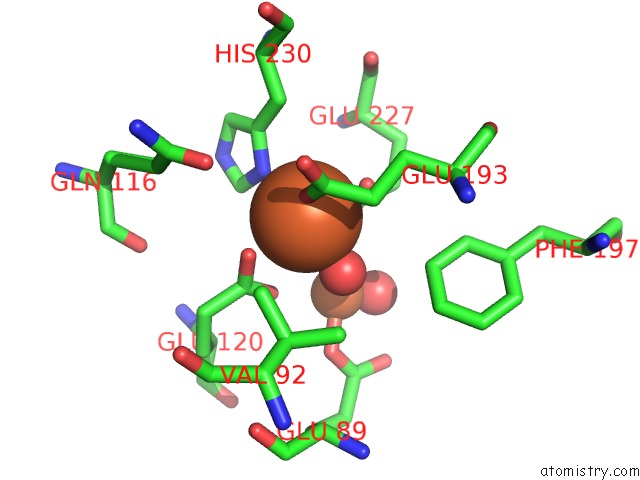
Mono view
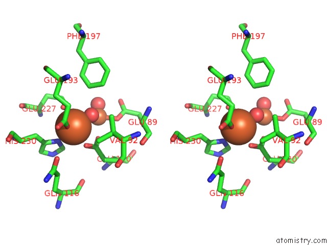
Stereo pair view
Mono view
Stereo pair view
A full contact list of Iron with other atoms in the Fe binding
site number 1 of Crystal Structure of the F127Y Mutant of Ribonucleotide Reductase R2 From Chlamydia Trachomatis within 5.0Å range:
|
Iron binding site 2 out of 2 in 2ani
Go back to
Iron binding site 2 out
of 2 in the Crystal Structure of the F127Y Mutant of Ribonucleotide Reductase R2 From Chlamydia Trachomatis
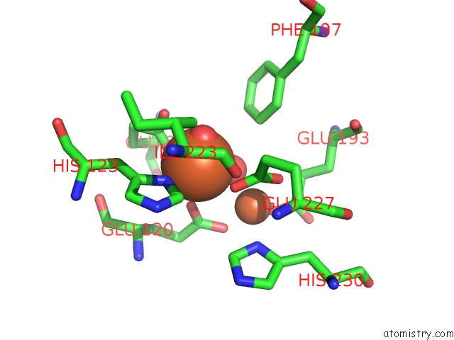
Mono view
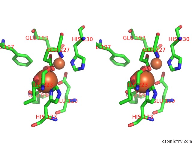
Stereo pair view
Mono view
Stereo pair view
A full contact list of Iron with other atoms in the Fe binding
site number 2 of Crystal Structure of the F127Y Mutant of Ribonucleotide Reductase R2 From Chlamydia Trachomatis within 5.0Å range:
|
Reference:
N.Voevodskaya,
M.Galander,
M.Hogbom,
P.Stenmark,
G.Mcclarty,
A.Graslund,
F.Lendzian.
Structure of the High-Valent Feiiifeiv State in Ribonucleotide Reductase (Rnr) of Chlamydia Trachomatis--Combined Epr, 57FE-, 1H-Endor and X-Ray Studies. Biochim.Biophys.Acta V.1774 1254 2007.
ISSN: ISSN 0006-3002
PubMed: 17827077
DOI: 10.1016/J.BBAPAP.2007.07.001
Page generated: Sat Aug 3 19:20:50 2024
ISSN: ISSN 0006-3002
PubMed: 17827077
DOI: 10.1016/J.BBAPAP.2007.07.001
Last articles
Zn in 9MJ5Zn in 9HNW
Zn in 9G0L
Zn in 9FNE
Zn in 9DZN
Zn in 9E0I
Zn in 9D32
Zn in 9DAK
Zn in 8ZXC
Zn in 8ZUF