Iron »
PDB 2aiu-2axx »
2at3 »
Iron in PDB 2at3: 1.00 A Crystal Structure of L123V/L133V Mutant of Nitrophorin 4 From Rhodnius Prolixus Complexed with Imidazole at pH 5.6
Protein crystallography data
The structure of 1.00 A Crystal Structure of L123V/L133V Mutant of Nitrophorin 4 From Rhodnius Prolixus Complexed with Imidazole at pH 5.6, PDB code: 2at3
was solved by
A.M.Amoia,
W.R.Montfort,
with X-Ray Crystallography technique. A brief refinement statistics is given in the table below:
| Resolution Low / High (Å) | 19.81 / 1.00 |
| Space group | C 1 2 1 |
| Cell size a, b, c (Å), α, β, γ (°) | 70.221, 42.743, 52.886, 90.00, 94.09, 90.00 |
| R / Rfree (%) | 14.4 / 16.7 |
Iron Binding Sites:
The binding sites of Iron atom in the 1.00 A Crystal Structure of L123V/L133V Mutant of Nitrophorin 4 From Rhodnius Prolixus Complexed with Imidazole at pH 5.6
(pdb code 2at3). This binding sites where shown within
5.0 Angstroms radius around Iron atom.
In total only one binding site of Iron was determined in the 1.00 A Crystal Structure of L123V/L133V Mutant of Nitrophorin 4 From Rhodnius Prolixus Complexed with Imidazole at pH 5.6, PDB code: 2at3:
In total only one binding site of Iron was determined in the 1.00 A Crystal Structure of L123V/L133V Mutant of Nitrophorin 4 From Rhodnius Prolixus Complexed with Imidazole at pH 5.6, PDB code: 2at3:
Iron binding site 1 out of 1 in 2at3
Go back to
Iron binding site 1 out
of 1 in the 1.00 A Crystal Structure of L123V/L133V Mutant of Nitrophorin 4 From Rhodnius Prolixus Complexed with Imidazole at pH 5.6
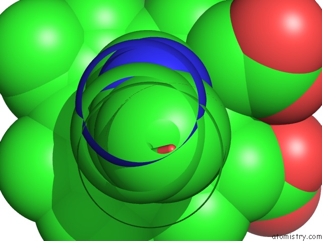
Mono view
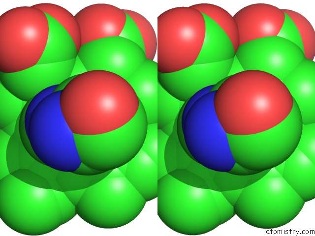
Stereo pair view
Mono view
Stereo pair view
A full contact list of Iron with other atoms in the Fe binding
site number 1 of 1.00 A Crystal Structure of L123V/L133V Mutant of Nitrophorin 4 From Rhodnius Prolixus Complexed with Imidazole at pH 5.6 within 5.0Å range:
|
Reference:
A.M.Amoia,
W.R.Montfort.
Heme Distortion in Nitrophorin 4: High Resolution Structures of Mutated Positions L123V and L133V and Heme Altered Proteins To Be Published.
Page generated: Sat Aug 3 19:22:42 2024
Last articles
Zn in 9JYWZn in 9IR4
Zn in 9IR3
Zn in 9GMX
Zn in 9GMW
Zn in 9JEJ
Zn in 9ERF
Zn in 9ERE
Zn in 9EGV
Zn in 9EGW