Iron »
PDB 3hnj-3i8r »
3i4y »
Iron in PDB 3i4y: Crystal Structure Determination of Catechol 1,2-Dioxygenase From Rhodococcus Opacus 1CP in Complex with 3,5-Dichlorocatechol
Enzymatic activity of Crystal Structure Determination of Catechol 1,2-Dioxygenase From Rhodococcus Opacus 1CP in Complex with 3,5-Dichlorocatechol
All present enzymatic activity of Crystal Structure Determination of Catechol 1,2-Dioxygenase From Rhodococcus Opacus 1CP in Complex with 3,5-Dichlorocatechol:
1.13.11.1;
1.13.11.1;
Protein crystallography data
The structure of Crystal Structure Determination of Catechol 1,2-Dioxygenase From Rhodococcus Opacus 1CP in Complex with 3,5-Dichlorocatechol, PDB code: 3i4y
was solved by
I.Matera,
M.Ferraroni,
M.Kolomytseva,
F.Briganti,
A.Scozzafava,
with X-Ray Crystallography technique. A brief refinement statistics is given in the table below:
| Resolution Low / High (Å) | 74.54 / 1.85 |
| Space group | C 1 2 1 |
| Cell size a, b, c (Å), α, β, γ (°) | 90.818, 37.619, 74.817, 90.00, 94.81, 90.00 |
| R / Rfree (%) | 20.4 / 26.7 |
Other elements in 3i4y:
The structure of Crystal Structure Determination of Catechol 1,2-Dioxygenase From Rhodococcus Opacus 1CP in Complex with 3,5-Dichlorocatechol also contains other interesting chemical elements:
| Chlorine | (Cl) | 4 atoms |
Iron Binding Sites:
The binding sites of Iron atom in the Crystal Structure Determination of Catechol 1,2-Dioxygenase From Rhodococcus Opacus 1CP in Complex with 3,5-Dichlorocatechol
(pdb code 3i4y). This binding sites where shown within
5.0 Angstroms radius around Iron atom.
In total only one binding site of Iron was determined in the Crystal Structure Determination of Catechol 1,2-Dioxygenase From Rhodococcus Opacus 1CP in Complex with 3,5-Dichlorocatechol, PDB code: 3i4y:
In total only one binding site of Iron was determined in the Crystal Structure Determination of Catechol 1,2-Dioxygenase From Rhodococcus Opacus 1CP in Complex with 3,5-Dichlorocatechol, PDB code: 3i4y:
Iron binding site 1 out of 1 in 3i4y
Go back to
Iron binding site 1 out
of 1 in the Crystal Structure Determination of Catechol 1,2-Dioxygenase From Rhodococcus Opacus 1CP in Complex with 3,5-Dichlorocatechol
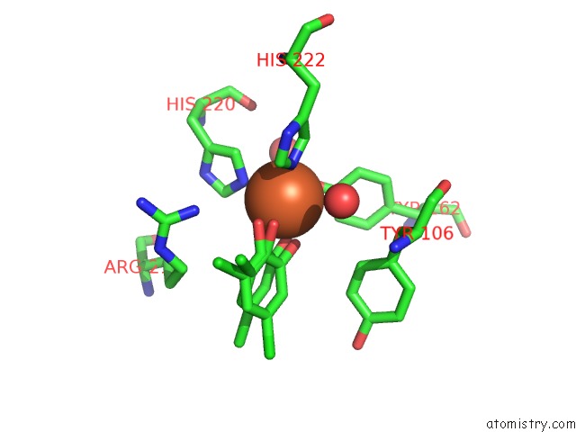
Mono view
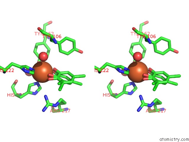
Stereo pair view
Mono view
Stereo pair view
A full contact list of Iron with other atoms in the Fe binding
site number 1 of Crystal Structure Determination of Catechol 1,2-Dioxygenase From Rhodococcus Opacus 1CP in Complex with 3,5-Dichlorocatechol within 5.0Å range:
|
Reference:
I.Matera,
M.Ferraroni,
M.Kolomytseva,
L.Golovleva,
A.Scozzafava,
F.Briganti.
Catechol 1,2-Dioxygenase From the Gram-Positive Rhodococcus Opacus 1CP: Quantitative Structure/Activity Relationship and the Crystal Structures of Native Enzyme and Catechols Adducts. J.Struct.Biol. V. 170 548 2010.
ISSN: ISSN 1047-8477
PubMed: 20040374
DOI: 10.1016/J.JSB.2009.12.023
Page generated: Tue Aug 5 02:10:36 2025
ISSN: ISSN 1047-8477
PubMed: 20040374
DOI: 10.1016/J.JSB.2009.12.023
Last articles
Fe in 3OGHFe in 3OIA
Fe in 3OFT
Fe in 3OGB
Fe in 3ODQ
Fe in 3OA8
Fe in 3OCD
Fe in 3ODB
Fe in 3OD7
Fe in 3O3M