Iron »
PDB 3p4r-3pcn »
3p6r »
Iron in PDB 3p6r: Crystal Structure of Cytochrome P450CAM Crystallized in the Presence of A Tethered Substrate Analog 3OH-ADAC1-Etg-Boc
Enzymatic activity of Crystal Structure of Cytochrome P450CAM Crystallized in the Presence of A Tethered Substrate Analog 3OH-ADAC1-Etg-Boc
All present enzymatic activity of Crystal Structure of Cytochrome P450CAM Crystallized in the Presence of A Tethered Substrate Analog 3OH-ADAC1-Etg-Boc:
1.14.15.1;
1.14.15.1;
Protein crystallography data
The structure of Crystal Structure of Cytochrome P450CAM Crystallized in the Presence of A Tethered Substrate Analog 3OH-ADAC1-Etg-Boc, PDB code: 3p6r
was solved by
Y.-T.Lee,
R.F.Wilson,
E.C.Glazer,
D.B.Goodin,
with X-Ray Crystallography technique. A brief refinement statistics is given in the table below:
| Resolution Low / High (Å) | 10.00 / 2.10 |
| Space group | P 21 21 21 |
| Cell size a, b, c (Å), α, β, γ (°) | 66.809, 75.008, 91.781, 90.00, 90.00, 90.00 |
| R / Rfree (%) | 21.7 / 24.6 |
Other elements in 3p6r:
The structure of Crystal Structure of Cytochrome P450CAM Crystallized in the Presence of A Tethered Substrate Analog 3OH-ADAC1-Etg-Boc also contains other interesting chemical elements:
| Chlorine | (Cl) | 1 atom |
Iron Binding Sites:
The binding sites of Iron atom in the Crystal Structure of Cytochrome P450CAM Crystallized in the Presence of A Tethered Substrate Analog 3OH-ADAC1-Etg-Boc
(pdb code 3p6r). This binding sites where shown within
5.0 Angstroms radius around Iron atom.
In total only one binding site of Iron was determined in the Crystal Structure of Cytochrome P450CAM Crystallized in the Presence of A Tethered Substrate Analog 3OH-ADAC1-Etg-Boc, PDB code: 3p6r:
In total only one binding site of Iron was determined in the Crystal Structure of Cytochrome P450CAM Crystallized in the Presence of A Tethered Substrate Analog 3OH-ADAC1-Etg-Boc, PDB code: 3p6r:
Iron binding site 1 out of 1 in 3p6r
Go back to
Iron binding site 1 out
of 1 in the Crystal Structure of Cytochrome P450CAM Crystallized in the Presence of A Tethered Substrate Analog 3OH-ADAC1-Etg-Boc
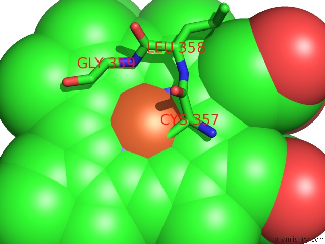
Mono view
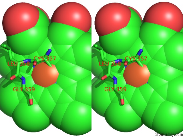
Stereo pair view
Mono view
Stereo pair view
A full contact list of Iron with other atoms in the Fe binding
site number 1 of Crystal Structure of Cytochrome P450CAM Crystallized in the Presence of A Tethered Substrate Analog 3OH-ADAC1-Etg-Boc within 5.0Å range:
|
Reference:
Y.-T.Lee,
E.C.Glazer,
R.F.Wilson,
D.B.Goodin.
Crystal Structure of Cytochrome P450CAM Crystallized in the Presence of A Tethered Substrate Analog 3OH-ADAC1-Etg-Boc To Be Published.
Page generated: Tue Aug 5 05:29:50 2025
Last articles
Fe in 3WFBFe in 3WCW
Fe in 3WCV
Fe in 3WEC
Fe in 3WCU
Fe in 3WCT
Fe in 3WCP
Fe in 3WCQ
Fe in 3WC8
Fe in 3WAH