Iron »
PDB 3p4r-3pcn »
3pcc »
Iron in PDB 3pcc: Structure of Protocatechuate 3,4-Dioxygenase Complexed with 4- Hydroxybenzoate
Enzymatic activity of Structure of Protocatechuate 3,4-Dioxygenase Complexed with 4- Hydroxybenzoate
All present enzymatic activity of Structure of Protocatechuate 3,4-Dioxygenase Complexed with 4- Hydroxybenzoate:
1.13.11.3;
1.13.11.3;
Protein crystallography data
The structure of Structure of Protocatechuate 3,4-Dioxygenase Complexed with 4- Hydroxybenzoate, PDB code: 3pcc
was solved by
N.Elango,
A.M.Orville,
J.D.Lipscomb,
D.H.Ohlendorf,
with X-Ray Crystallography technique. A brief refinement statistics is given in the table below:
| Resolution Low / High (Å) | 6.00 / 1.98 |
| Space group | I 1 2 1 |
| Cell size a, b, c (Å), α, β, γ (°) | 197.410, 127.970, 134.880, 90.00, 97.80, 90.00 |
| R / Rfree (%) | n/a / n/a |
Iron Binding Sites:
The binding sites of Iron atom in the Structure of Protocatechuate 3,4-Dioxygenase Complexed with 4- Hydroxybenzoate
(pdb code 3pcc). This binding sites where shown within
5.0 Angstroms radius around Iron atom.
In total 6 binding sites of Iron where determined in the Structure of Protocatechuate 3,4-Dioxygenase Complexed with 4- Hydroxybenzoate, PDB code: 3pcc:
Jump to Iron binding site number: 1; 2; 3; 4; 5; 6;
In total 6 binding sites of Iron where determined in the Structure of Protocatechuate 3,4-Dioxygenase Complexed with 4- Hydroxybenzoate, PDB code: 3pcc:
Jump to Iron binding site number: 1; 2; 3; 4; 5; 6;
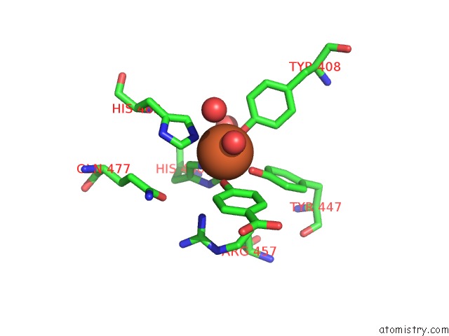
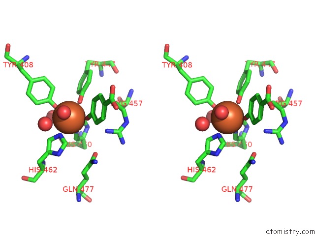
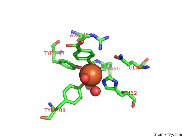
Iron binding site 1 out of 6 in 3pcc
Go back to
Iron binding site 1 out
of 6 in the Structure of Protocatechuate 3,4-Dioxygenase Complexed with 4- Hydroxybenzoate
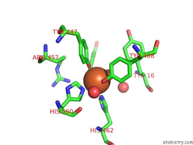
Mono view
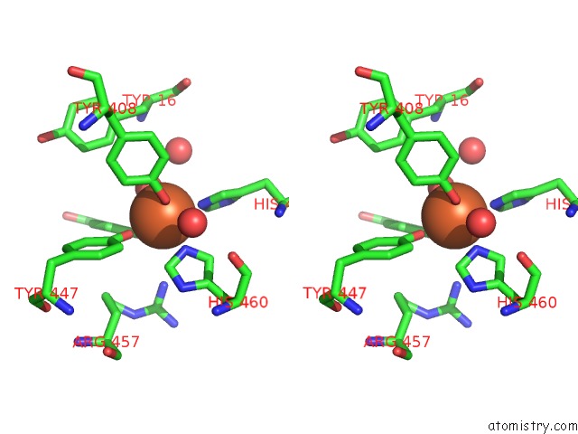
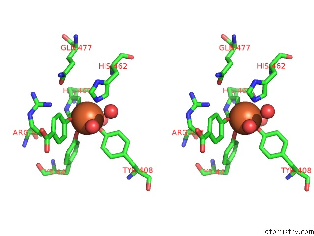
Stereo pair view
Mono view
Stereo pair view
A full contact list of Iron with other atoms in the Fe binding
site number 1 of Structure of Protocatechuate 3,4-Dioxygenase Complexed with 4- Hydroxybenzoate within 5.0Å range:
|
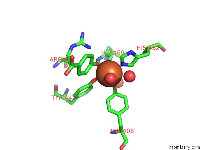
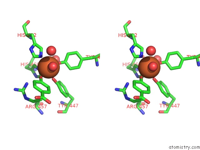
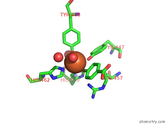
Iron binding site 2 out of 6 in 3pcc
Go back to
Iron binding site 2 out
of 6 in the Structure of Protocatechuate 3,4-Dioxygenase Complexed with 4- Hydroxybenzoate
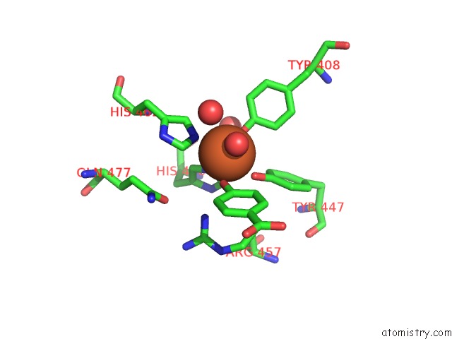
Mono view
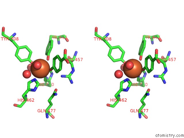
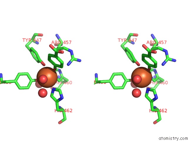
Stereo pair view
Mono view
Stereo pair view
A full contact list of Iron with other atoms in the Fe binding
site number 2 of Structure of Protocatechuate 3,4-Dioxygenase Complexed with 4- Hydroxybenzoate within 5.0Å range:
|
Iron binding site 3 out of 6 in 3pcc
Go back to
Iron binding site 3 out
of 6 in the Structure of Protocatechuate 3,4-Dioxygenase Complexed with 4- Hydroxybenzoate
Mono view
Stereo pair view
Mono view
Stereo pair view
A full contact list of Iron with other atoms in the Fe binding
site number 3 of Structure of Protocatechuate 3,4-Dioxygenase Complexed with 4- Hydroxybenzoate within 5.0Å range:
|
Iron binding site 4 out of 6 in 3pcc
Go back to
Iron binding site 4 out
of 6 in the Structure of Protocatechuate 3,4-Dioxygenase Complexed with 4- Hydroxybenzoate
Mono view
Stereo pair view
Mono view
Stereo pair view
A full contact list of Iron with other atoms in the Fe binding
site number 4 of Structure of Protocatechuate 3,4-Dioxygenase Complexed with 4- Hydroxybenzoate within 5.0Å range:
|
Iron binding site 5 out of 6 in 3pcc
Go back to
Iron binding site 5 out
of 6 in the Structure of Protocatechuate 3,4-Dioxygenase Complexed with 4- Hydroxybenzoate
Mono view
Stereo pair view
Mono view
Stereo pair view
A full contact list of Iron with other atoms in the Fe binding
site number 5 of Structure of Protocatechuate 3,4-Dioxygenase Complexed with 4- Hydroxybenzoate within 5.0Å range:
|
Iron binding site 6 out of 6 in 3pcc
Go back to
Iron binding site 6 out
of 6 in the Structure of Protocatechuate 3,4-Dioxygenase Complexed with 4- Hydroxybenzoate
Mono view
Stereo pair view
Mono view
Stereo pair view
A full contact list of Iron with other atoms in the Fe binding
site number 6 of Structure of Protocatechuate 3,4-Dioxygenase Complexed with 4- Hydroxybenzoate within 5.0Å range:
|
Reference:
A.M.Orville,
N.Elango,
J.D.Lipscomb,
D.H.Ohlendorf.
Structures of Competitive Inhibitor Complexes of Protocatechuate 3,4-Dioxygenase: Multiple Exogenous Ligand Binding Orientations Within the Active Site. Biochemistry V. 36 10039 1997.
ISSN: ISSN 0006-2960
PubMed: 9254599
DOI: 10.1021/BI970468N
Page generated: Tue Aug 5 05:37:03 2025
ISSN: ISSN 0006-2960
PubMed: 9254599
DOI: 10.1021/BI970468N
Last articles
Fe in 3WFBFe in 3WCW
Fe in 3WCV
Fe in 3WEC
Fe in 3WCU
Fe in 3WCT
Fe in 3WCP
Fe in 3WCQ
Fe in 3WC8
Fe in 3WAH