Iron »
PDB 3pcq-3puq »
3pl1 »
Iron in PDB 3pl1: Determination of the Crystal Structure of the Pyrazinamidase From M.Tuberculosis : A Structure-Function Analysis For Prediction Resistance to Pyrazinamide.
Enzymatic activity of Determination of the Crystal Structure of the Pyrazinamidase From M.Tuberculosis : A Structure-Function Analysis For Prediction Resistance to Pyrazinamide.
All present enzymatic activity of Determination of the Crystal Structure of the Pyrazinamidase From M.Tuberculosis : A Structure-Function Analysis For Prediction Resistance to Pyrazinamide.:
3.5.1.19;
3.5.1.19;
Protein crystallography data
The structure of Determination of the Crystal Structure of the Pyrazinamidase From M.Tuberculosis : A Structure-Function Analysis For Prediction Resistance to Pyrazinamide., PDB code: 3pl1
was solved by
S.Petrella,
N.Gelus-Ziental,
C.Mayer,
W.Sougakoff,
with X-Ray Crystallography technique. A brief refinement statistics is given in the table below:
| Resolution Low / High (Å) | 42.03 / 2.20 |
| Space group | P 61 2 2 |
| Cell size a, b, c (Å), α, β, γ (°) | 84.100, 84.100, 100.000, 90.00, 90.00, 120.00 |
| R / Rfree (%) | 19.5 / 24 |
Iron Binding Sites:
The binding sites of Iron atom in the Determination of the Crystal Structure of the Pyrazinamidase From M.Tuberculosis : A Structure-Function Analysis For Prediction Resistance to Pyrazinamide.
(pdb code 3pl1). This binding sites where shown within
5.0 Angstroms radius around Iron atom.
In total only one binding site of Iron was determined in the Determination of the Crystal Structure of the Pyrazinamidase From M.Tuberculosis : A Structure-Function Analysis For Prediction Resistance to Pyrazinamide., PDB code: 3pl1:
In total only one binding site of Iron was determined in the Determination of the Crystal Structure of the Pyrazinamidase From M.Tuberculosis : A Structure-Function Analysis For Prediction Resistance to Pyrazinamide., PDB code: 3pl1:
Iron binding site 1 out of 1 in 3pl1
Go back to
Iron binding site 1 out
of 1 in the Determination of the Crystal Structure of the Pyrazinamidase From M.Tuberculosis : A Structure-Function Analysis For Prediction Resistance to Pyrazinamide.
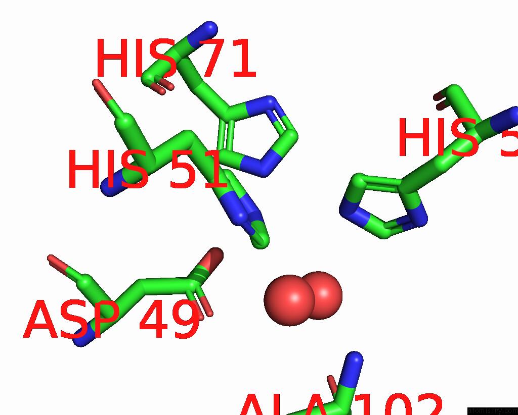
Mono view
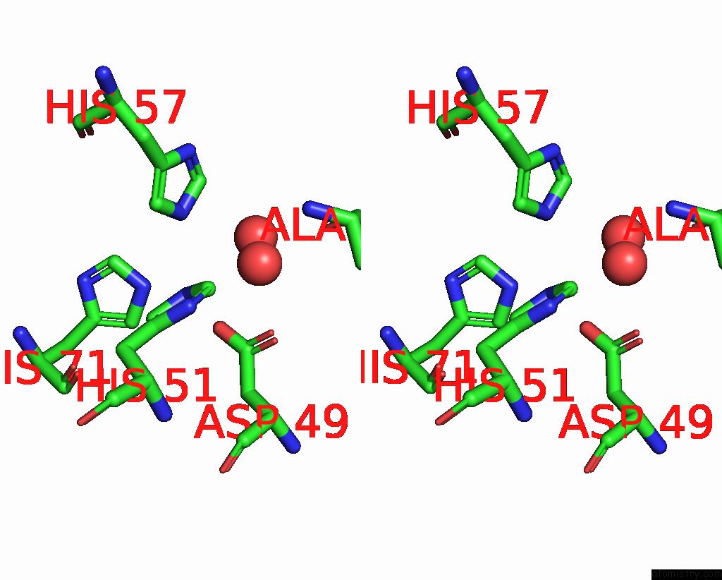
Stereo pair view
Mono view
Stereo pair view
A full contact list of Iron with other atoms in the Fe binding
site number 1 of Determination of the Crystal Structure of the Pyrazinamidase From M.Tuberculosis : A Structure-Function Analysis For Prediction Resistance to Pyrazinamide. within 5.0Å range:
|
Reference:
S.Petrella,
N.Gelus-Ziental,
A.Maudry,
C.Laurans,
R.Boudjelloul,
W.Sougakoff.
Crystal Structure of the Pyrazinamidase of Mycobacterium Tuberculosis: Insights Into Natural and Acquired Resistance to Pyrazinamide. Plos One V. 6 15785 2011.
ISSN: ESSN 1932-6203
PubMed: 21283666
DOI: 10.1371/JOURNAL.PONE.0015785
Page generated: Tue Aug 5 05:45:37 2025
ISSN: ESSN 1932-6203
PubMed: 21283666
DOI: 10.1371/JOURNAL.PONE.0015785
Last articles
Fe in 3WFBFe in 3WCW
Fe in 3WCV
Fe in 3WEC
Fe in 3WCU
Fe in 3WCT
Fe in 3WCP
Fe in 3WCQ
Fe in 3WC8
Fe in 3WAH