Iron »
PDB 3uhg-3v2z »
3uk4 »
Iron in PDB 3uk4: Crystal Structure of C-Lobe of Bovine Lactoferrin Complexed with 1,2, 5-Pentanetriol at 1.98 A Resolution
Protein crystallography data
The structure of Crystal Structure of C-Lobe of Bovine Lactoferrin Complexed with 1,2, 5-Pentanetriol at 1.98 A Resolution, PDB code: 3uk4
was solved by
P.K.Shukla,
L.Gautam,
M.Sinha,
P.Kaur,
S.Sharma,
T.P.Singh,
with X-Ray Crystallography technique. A brief refinement statistics is given in the table below:
| Resolution Low / High (Å) | 62.80 / 1.98 |
| Space group | P 1 21 1 |
| Cell size a, b, c (Å), α, β, γ (°) | 62.925, 50.215, 65.633, 90.00, 106.88, 90.00 |
| R / Rfree (%) | 19.8 / 24.1 |
Other elements in 3uk4:
The structure of Crystal Structure of C-Lobe of Bovine Lactoferrin Complexed with 1,2, 5-Pentanetriol at 1.98 A Resolution also contains other interesting chemical elements:
| Zinc | (Zn) | 2 atoms |
Iron Binding Sites:
The binding sites of Iron atom in the Crystal Structure of C-Lobe of Bovine Lactoferrin Complexed with 1,2, 5-Pentanetriol at 1.98 A Resolution
(pdb code 3uk4). This binding sites where shown within
5.0 Angstroms radius around Iron atom.
In total only one binding site of Iron was determined in the Crystal Structure of C-Lobe of Bovine Lactoferrin Complexed with 1,2, 5-Pentanetriol at 1.98 A Resolution, PDB code: 3uk4:
In total only one binding site of Iron was determined in the Crystal Structure of C-Lobe of Bovine Lactoferrin Complexed with 1,2, 5-Pentanetriol at 1.98 A Resolution, PDB code: 3uk4:
Iron binding site 1 out of 1 in 3uk4
Go back to
Iron binding site 1 out
of 1 in the Crystal Structure of C-Lobe of Bovine Lactoferrin Complexed with 1,2, 5-Pentanetriol at 1.98 A Resolution
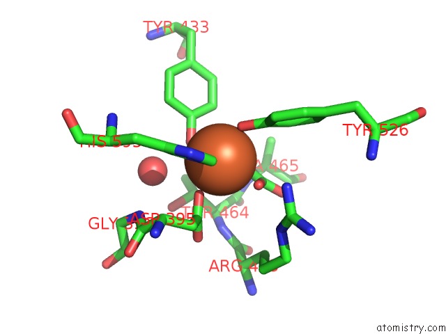
Mono view
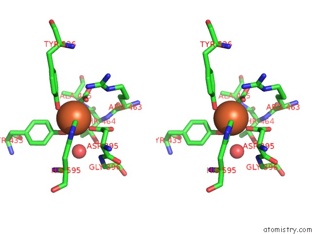
Stereo pair view
Mono view
Stereo pair view
A full contact list of Iron with other atoms in the Fe binding
site number 1 of Crystal Structure of C-Lobe of Bovine Lactoferrin Complexed with 1,2, 5-Pentanetriol at 1.98 A Resolution within 5.0Å range:
|
Reference:
P.K.Shukla,
L.Gautam,
M.Sinha,
P.Kaur,
S.Sharma,
T.P.Singh.
Crystal Structure of C-Lobe of Bovine Lactoferrin Complexed with 1,2,5-Pentanetriol at 1.98 A Resolution To Be Published.
Page generated: Tue Aug 5 07:28:43 2025
Last articles
Fe in 4DJAFe in 4DIK
Fe in 4DIL
Fe in 4DIG
Fe in 4DHV
Fe in 4DHL
Fe in 4DI0
Fe in 4DCX
Fe in 4DCY
Fe in 4D8G