Iron »
PDB 3vtj-3wg7 »
3vv9 »
Iron in PDB 3vv9: Crystal Structure of Cyanide-Insensitive Alternative Oxidase From Trypanosoma Brucei
Protein crystallography data
The structure of Crystal Structure of Cyanide-Insensitive Alternative Oxidase From Trypanosoma Brucei, PDB code: 3vv9
was solved by
T.Shiba,
Y.Kido,
K.Sakamoto,
D.K.Inaoka,
C.Tsuge,
R.Tatsumi,
E.O.Balogun,
T.Nara,
T.Aoki,
T.Honma,
A.Tanaka,
M.Inoue,
S.Matsuoka,
H.Saimoto,
A.L.Moore,
S.Harada,
K.Kita,
with X-Ray Crystallography technique. A brief refinement statistics is given in the table below:
| Resolution Low / High (Å) | 45.38 / 2.85 |
| Space group | C 1 2 1 |
| Cell size a, b, c (Å), α, β, γ (°) | 261.312, 63.120, 136.457, 90.00, 121.38, 90.00 |
| R / Rfree (%) | 19.2 / 24.7 |
Iron Binding Sites:
The binding sites of Iron atom in the Crystal Structure of Cyanide-Insensitive Alternative Oxidase From Trypanosoma Brucei
(pdb code 3vv9). This binding sites where shown within
5.0 Angstroms radius around Iron atom.
In total 8 binding sites of Iron where determined in the Crystal Structure of Cyanide-Insensitive Alternative Oxidase From Trypanosoma Brucei, PDB code: 3vv9:
Jump to Iron binding site number: 1; 2; 3; 4; 5; 6; 7; 8;
In total 8 binding sites of Iron where determined in the Crystal Structure of Cyanide-Insensitive Alternative Oxidase From Trypanosoma Brucei, PDB code: 3vv9:
Jump to Iron binding site number: 1; 2; 3; 4; 5; 6; 7; 8;
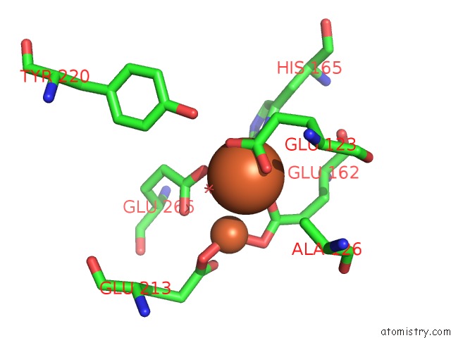
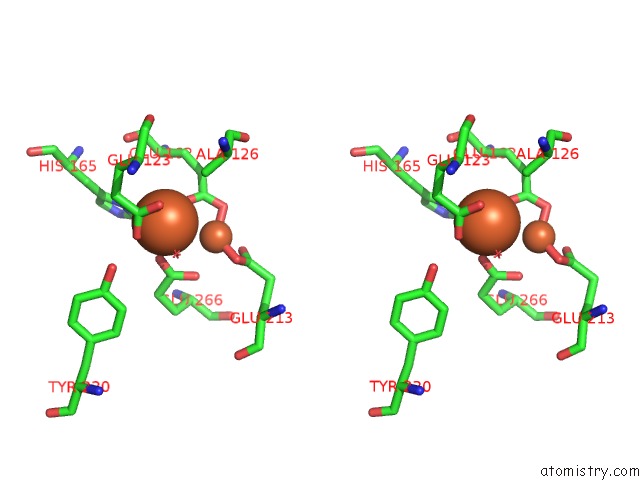
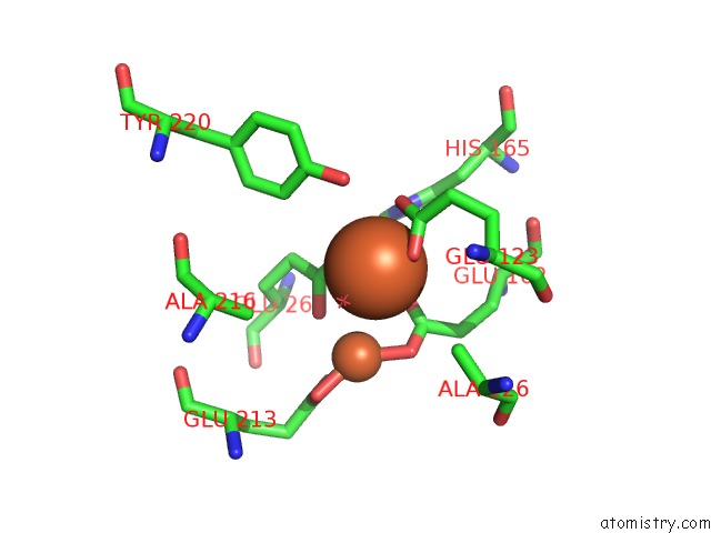
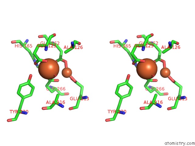
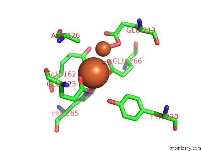
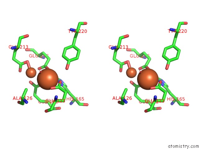
Iron binding site 1 out of 8 in 3vv9
Go back to
Iron binding site 1 out
of 8 in the Crystal Structure of Cyanide-Insensitive Alternative Oxidase From Trypanosoma Brucei
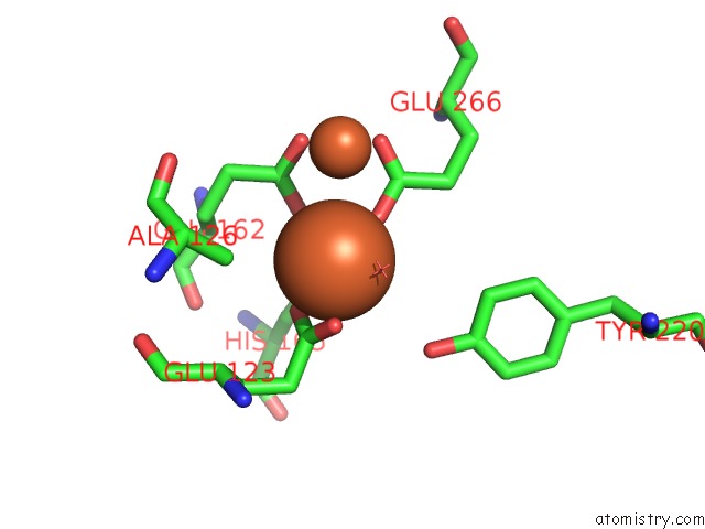
Mono view
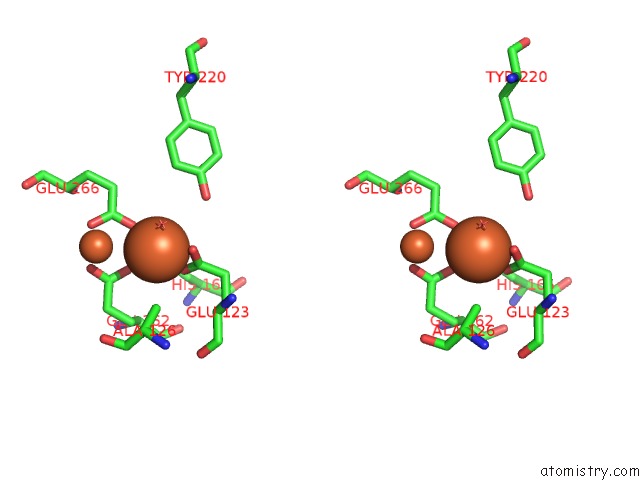
Stereo pair view
Mono view
Stereo pair view
A full contact list of Iron with other atoms in the Fe binding
site number 1 of Crystal Structure of Cyanide-Insensitive Alternative Oxidase From Trypanosoma Brucei within 5.0Å range:
|
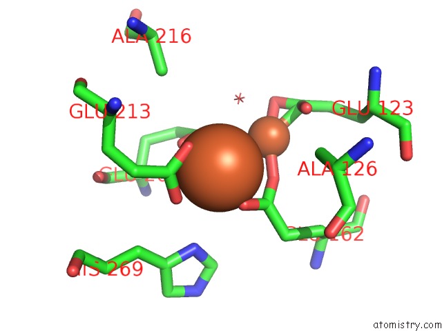
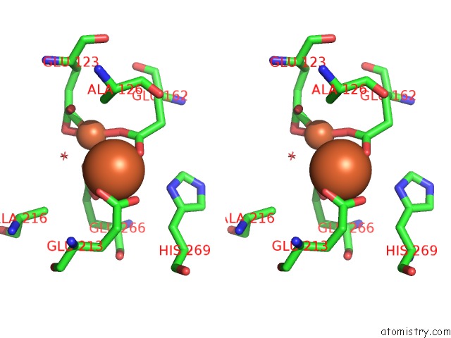
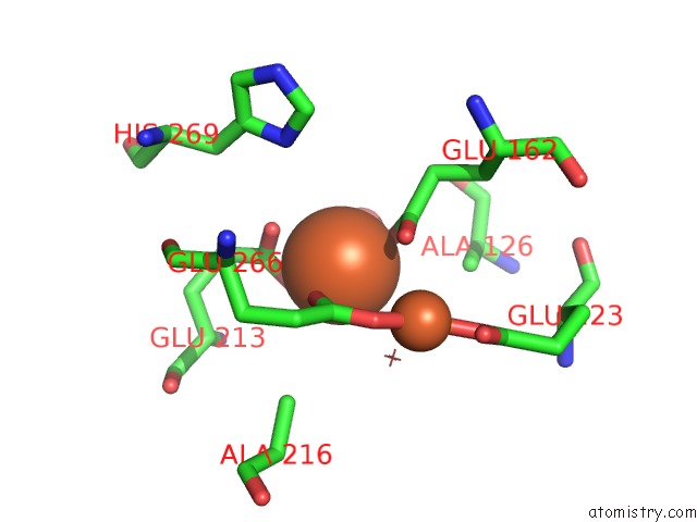
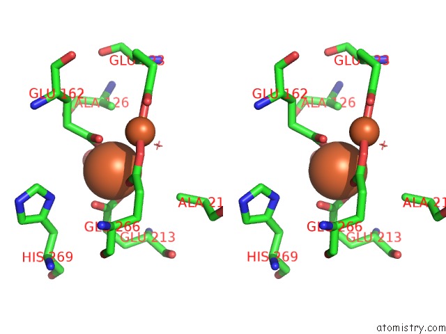
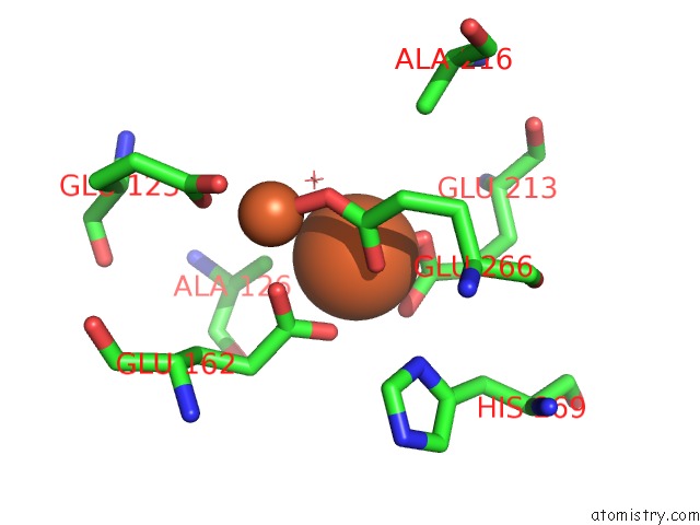
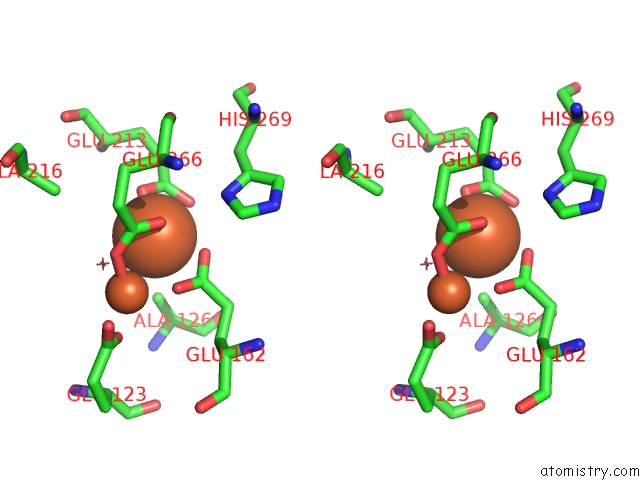
Iron binding site 2 out of 8 in 3vv9
Go back to
Iron binding site 2 out
of 8 in the Crystal Structure of Cyanide-Insensitive Alternative Oxidase From Trypanosoma Brucei
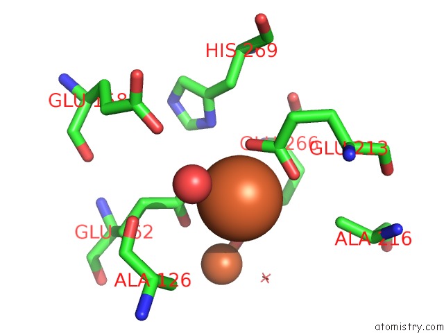
Mono view
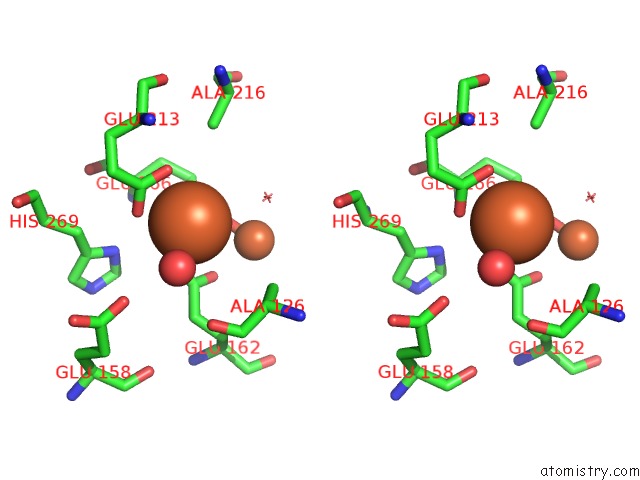
Stereo pair view
Mono view
Stereo pair view
A full contact list of Iron with other atoms in the Fe binding
site number 2 of Crystal Structure of Cyanide-Insensitive Alternative Oxidase From Trypanosoma Brucei within 5.0Å range:
|
Iron binding site 3 out of 8 in 3vv9
Go back to
Iron binding site 3 out
of 8 in the Crystal Structure of Cyanide-Insensitive Alternative Oxidase From Trypanosoma Brucei
Mono view
Stereo pair view
Mono view
Stereo pair view
A full contact list of Iron with other atoms in the Fe binding
site number 3 of Crystal Structure of Cyanide-Insensitive Alternative Oxidase From Trypanosoma Brucei within 5.0Å range:
|
Iron binding site 4 out of 8 in 3vv9
Go back to
Iron binding site 4 out
of 8 in the Crystal Structure of Cyanide-Insensitive Alternative Oxidase From Trypanosoma Brucei
Mono view
Stereo pair view
Mono view
Stereo pair view
A full contact list of Iron with other atoms in the Fe binding
site number 4 of Crystal Structure of Cyanide-Insensitive Alternative Oxidase From Trypanosoma Brucei within 5.0Å range:
|
Iron binding site 5 out of 8 in 3vv9
Go back to
Iron binding site 5 out
of 8 in the Crystal Structure of Cyanide-Insensitive Alternative Oxidase From Trypanosoma Brucei
Mono view
Stereo pair view
Mono view
Stereo pair view
A full contact list of Iron with other atoms in the Fe binding
site number 5 of Crystal Structure of Cyanide-Insensitive Alternative Oxidase From Trypanosoma Brucei within 5.0Å range:
|
Iron binding site 6 out of 8 in 3vv9
Go back to
Iron binding site 6 out
of 8 in the Crystal Structure of Cyanide-Insensitive Alternative Oxidase From Trypanosoma Brucei
Mono view
Stereo pair view
Mono view
Stereo pair view
A full contact list of Iron with other atoms in the Fe binding
site number 6 of Crystal Structure of Cyanide-Insensitive Alternative Oxidase From Trypanosoma Brucei within 5.0Å range:
|
Iron binding site 7 out of 8 in 3vv9
Go back to
Iron binding site 7 out
of 8 in the Crystal Structure of Cyanide-Insensitive Alternative Oxidase From Trypanosoma Brucei
Mono view
Stereo pair view
Mono view
Stereo pair view
A full contact list of Iron with other atoms in the Fe binding
site number 7 of Crystal Structure of Cyanide-Insensitive Alternative Oxidase From Trypanosoma Brucei within 5.0Å range:
|
Iron binding site 8 out of 8 in 3vv9
Go back to
Iron binding site 8 out
of 8 in the Crystal Structure of Cyanide-Insensitive Alternative Oxidase From Trypanosoma Brucei
Mono view
Stereo pair view
Mono view
Stereo pair view
A full contact list of Iron with other atoms in the Fe binding
site number 8 of Crystal Structure of Cyanide-Insensitive Alternative Oxidase From Trypanosoma Brucei within 5.0Å range:
|
Reference:
T.Shiba,
Y.Kido,
K.Sakamoto,
D.K.Inaoka,
C.Tsuge,
R.Tatsumi,
G.Takahashi,
E.O.Balogun,
T.Nara,
T.Aoki,
T.Honma,
A.Tanaka,
M.Inoue,
S.Matsuoka,
H.Saimoto,
A.L.Moore,
S.Harada,
K.Kita.
Structure of the Trypanosome Cyanide-Insensitive Alternative Oxidase Proc.Natl.Acad.Sci.Usa V. 110 4580 2013.
ISSN: ISSN 0027-8424
PubMed: 23487766
DOI: 10.1073/PNAS.1218386110
Page generated: Sun Aug 4 22:26:58 2024
ISSN: ISSN 0027-8424
PubMed: 23487766
DOI: 10.1073/PNAS.1218386110
Last articles
Zn in 9J0NZn in 9J0O
Zn in 9J0P
Zn in 9FJX
Zn in 9EKB
Zn in 9C0F
Zn in 9CAH
Zn in 9CH0
Zn in 9CH3
Zn in 9CH1