Iron »
PDB 3vtj-3wg7 »
3w1w »
Iron in PDB 3w1w: Protein-Drug Complex
Enzymatic activity of Protein-Drug Complex
All present enzymatic activity of Protein-Drug Complex:
4.99.1.1;
4.99.1.1;
Protein crystallography data
The structure of Protein-Drug Complex, PDB code: 3w1w
was solved by
R.Ishii,
V.Gupta,
Y.Yamaguchi,
H.Handa,
O.Nureki,
with X-Ray Crystallography technique. A brief refinement statistics is given in the table below:
| Resolution Low / High (Å) | 34.28 / 2.01 |
| Space group | P 21 21 21 |
| Cell size a, b, c (Å), α, β, γ (°) | 87.531, 93.627, 110.287, 90.00, 90.00, 90.00 |
| R / Rfree (%) | 19.1 / 22.9 |
Iron Binding Sites:
The binding sites of Iron atom in the Protein-Drug Complex
(pdb code 3w1w). This binding sites where shown within
5.0 Angstroms radius around Iron atom.
In total 4 binding sites of Iron where determined in the Protein-Drug Complex, PDB code: 3w1w:
Jump to Iron binding site number: 1; 2; 3; 4;
In total 4 binding sites of Iron where determined in the Protein-Drug Complex, PDB code: 3w1w:
Jump to Iron binding site number: 1; 2; 3; 4;
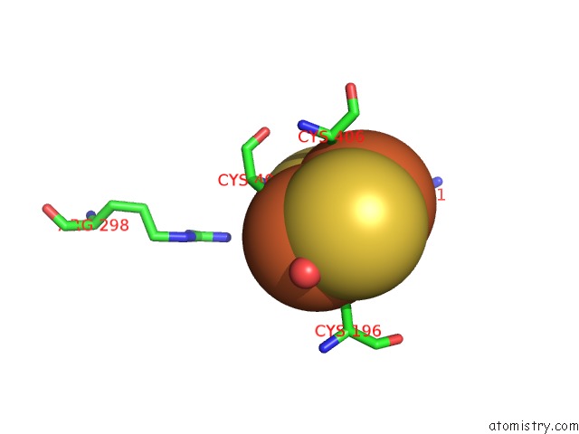
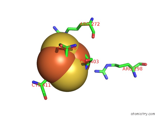
Iron binding site 1 out of 4 in 3w1w
Go back to
Iron binding site 1 out
of 4 in the Protein-Drug Complex
Mono view
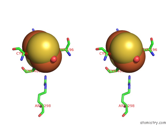
Stereo pair view
Mono view
Stereo pair view
A full contact list of Iron with other atoms in the Fe binding
site number 1 of Protein-Drug Complex within 5.0Å range:
|
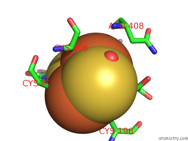
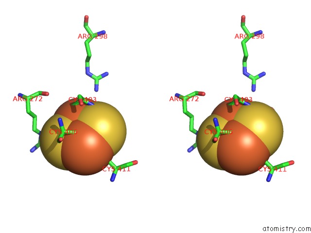
Iron binding site 2 out of 4 in 3w1w
Go back to
Iron binding site 2 out
of 4 in the Protein-Drug Complex
Mono view
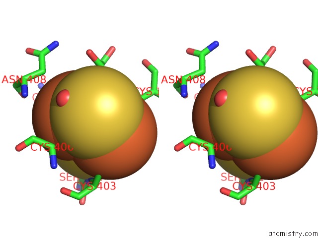
Stereo pair view
Mono view
Stereo pair view
A full contact list of Iron with other atoms in the Fe binding
site number 2 of Protein-Drug Complex within 5.0Å range:
|
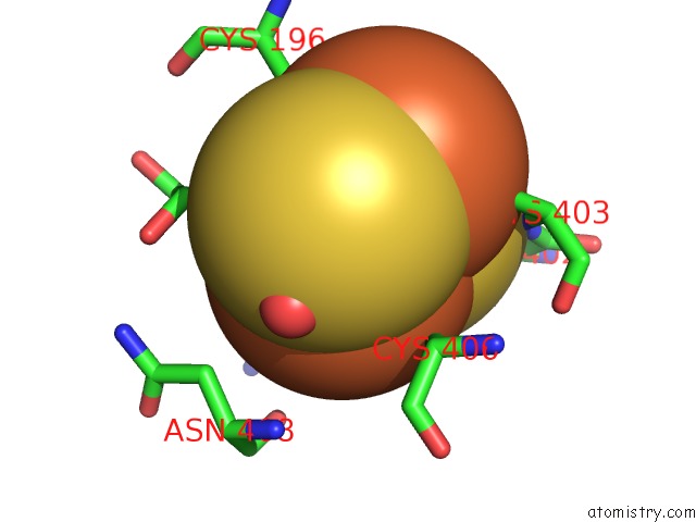
Iron binding site 3 out of 4 in 3w1w
Go back to
Iron binding site 3 out
of 4 in the Protein-Drug Complex
Mono view
Stereo pair view
Mono view
Stereo pair view
A full contact list of Iron with other atoms in the Fe binding
site number 3 of Protein-Drug Complex within 5.0Å range:
|
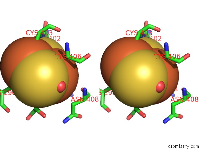
Iron binding site 4 out of 4 in 3w1w
Go back to
Iron binding site 4 out
of 4 in the Protein-Drug Complex
Mono view
Stereo pair view
Mono view
Stereo pair view
A full contact list of Iron with other atoms in the Fe binding
site number 4 of Protein-Drug Complex within 5.0Å range:
|
Reference:
V.Gupta,
S.Liu,
H.Ando,
R.Ishii,
S.Tateno,
Y.Kaneko,
M.Yugami,
S.Sakamoto,
Y.Yamaguchi,
O.Nureki,
H.Handa.
Salicylic Acid Induces Mitochondrial Injury By Inhibiting Ferrochelatase Heme Biosynthesis Activity Mol.Pharmacol. V. 84 824 2013.
ISSN: ISSN 0026-895X
PubMed: 24043703
DOI: 10.1124/MOL.113.087940
Page generated: Sun Aug 4 22:28:20 2024
ISSN: ISSN 0026-895X
PubMed: 24043703
DOI: 10.1124/MOL.113.087940
Last articles
Zn in 9J0NZn in 9J0O
Zn in 9J0P
Zn in 9FJX
Zn in 9EKB
Zn in 9C0F
Zn in 9CAH
Zn in 9CH0
Zn in 9CH3
Zn in 9CH1