Iron »
PDB 3vtj-3wg7 »
3w9c »
Iron in PDB 3w9c: Crystal Structure of the Electron Transfer Complex of Cytochrome P450CAM with Putidaredoxin
Enzymatic activity of Crystal Structure of the Electron Transfer Complex of Cytochrome P450CAM with Putidaredoxin
All present enzymatic activity of Crystal Structure of the Electron Transfer Complex of Cytochrome P450CAM with Putidaredoxin:
1.14.15.1;
1.14.15.1;
Protein crystallography data
The structure of Crystal Structure of the Electron Transfer Complex of Cytochrome P450CAM with Putidaredoxin, PDB code: 3w9c
was solved by
Y.Kikui,
Y.Hiruma,
M.A.Hass,
H.Koteishi,
M.Ubbink,
M.Nojiri,
with X-Ray Crystallography technique. A brief refinement statistics is given in the table below:
| Resolution Low / High (Å) | 61.78 / 2.50 |
| Space group | C 1 2 1 |
| Cell size a, b, c (Å), α, β, γ (°) | 101.717, 77.991, 60.016, 90.00, 95.57, 90.00 |
| R / Rfree (%) | 18.5 / 25.1 |
Iron Binding Sites:
The binding sites of Iron atom in the Crystal Structure of the Electron Transfer Complex of Cytochrome P450CAM with Putidaredoxin
(pdb code 3w9c). This binding sites where shown within
5.0 Angstroms radius around Iron atom.
In total 3 binding sites of Iron where determined in the Crystal Structure of the Electron Transfer Complex of Cytochrome P450CAM with Putidaredoxin, PDB code: 3w9c:
Jump to Iron binding site number: 1; 2; 3;
In total 3 binding sites of Iron where determined in the Crystal Structure of the Electron Transfer Complex of Cytochrome P450CAM with Putidaredoxin, PDB code: 3w9c:
Jump to Iron binding site number: 1; 2; 3;
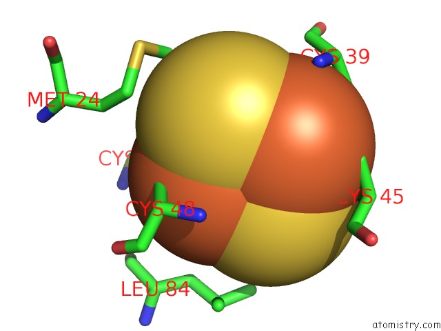
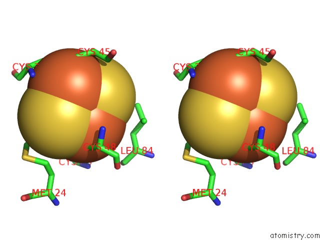
Iron binding site 1 out of 3 in 3w9c
Go back to
Iron binding site 1 out
of 3 in the Crystal Structure of the Electron Transfer Complex of Cytochrome P450CAM with Putidaredoxin
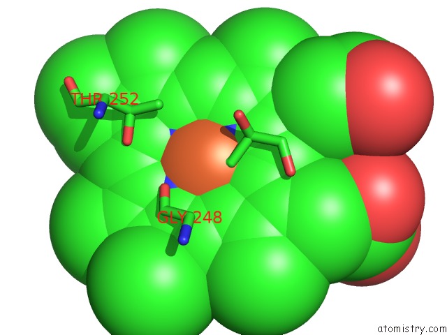
Mono view
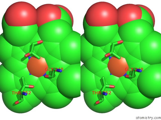
Stereo pair view
Mono view
Stereo pair view
A full contact list of Iron with other atoms in the Fe binding
site number 1 of Crystal Structure of the Electron Transfer Complex of Cytochrome P450CAM with Putidaredoxin within 5.0Å range:
|
Iron binding site 2 out of 3 in 3w9c
Go back to
Iron binding site 2 out
of 3 in the Crystal Structure of the Electron Transfer Complex of Cytochrome P450CAM with Putidaredoxin
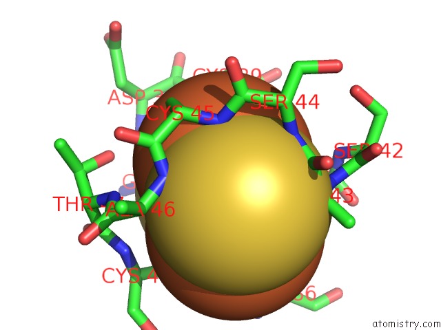
Mono view
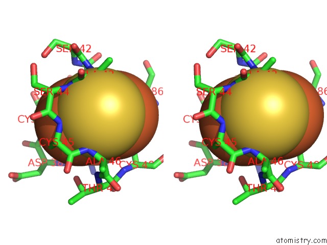
Stereo pair view
Mono view
Stereo pair view
A full contact list of Iron with other atoms in the Fe binding
site number 2 of Crystal Structure of the Electron Transfer Complex of Cytochrome P450CAM with Putidaredoxin within 5.0Å range:
|
Iron binding site 3 out of 3 in 3w9c
Go back to
Iron binding site 3 out
of 3 in the Crystal Structure of the Electron Transfer Complex of Cytochrome P450CAM with Putidaredoxin
Mono view
Stereo pair view
Mono view
Stereo pair view
A full contact list of Iron with other atoms in the Fe binding
site number 3 of Crystal Structure of the Electron Transfer Complex of Cytochrome P450CAM with Putidaredoxin within 5.0Å range:
|
Reference:
Y.Hiruma,
M.A.Hass,
Y.Kikui,
W.M.Liu,
B.Olmez,
S.P.Skinner,
A.Blok,
A.Kloosterman,
H.Koteishi,
F.Lohr,
H.Schwalbe,
M.Nojiri,
M.Ubbink.
The Structure of the Cytochrome P450CAM-Putidaredoxin Complex Determined By Paramagnetic uc(Nmr) Spectroscopy and Crystallography. J.Mol.Biol. 2013.
ISSN: ESSN 1089-8638
PubMed: 23856620
DOI: 10.1016/J.JMB.2013.07.006
Page generated: Sun Aug 4 22:32:24 2024
ISSN: ESSN 1089-8638
PubMed: 23856620
DOI: 10.1016/J.JMB.2013.07.006
Last articles
Zn in 9J0NZn in 9J0O
Zn in 9J0P
Zn in 9FJX
Zn in 9EKB
Zn in 9C0F
Zn in 9CAH
Zn in 9CH0
Zn in 9CH3
Zn in 9CH1