Iron »
PDB 3vtj-3wg7 »
3wcq »
Iron in PDB 3wcq: Crystal Structure Analysis of Cyanidioschyzon Melorae Ferredoxin D58N Mutant
Protein crystallography data
The structure of Crystal Structure Analysis of Cyanidioschyzon Melorae Ferredoxin D58N Mutant, PDB code: 3wcq
was solved by
Y.Ueno,
T.Matsumoto,
A.Yamano,
T.Imai,
Y.Morimoto,
with X-Ray Crystallography technique. A brief refinement statistics is given in the table below:
| Resolution Low / High (Å) | 26.97 / 0.97 |
| Space group | P 21 21 21 |
| Cell size a, b, c (Å), α, β, γ (°) | 34.026, 46.527, 66.139, 90.00, 90.00, 90.00 |
| R / Rfree (%) | 15.1 / 17 |
Iron Binding Sites:
The binding sites of Iron atom in the Crystal Structure Analysis of Cyanidioschyzon Melorae Ferredoxin D58N Mutant
(pdb code 3wcq). This binding sites where shown within
5.0 Angstroms radius around Iron atom.
In total 2 binding sites of Iron where determined in the Crystal Structure Analysis of Cyanidioschyzon Melorae Ferredoxin D58N Mutant, PDB code: 3wcq:
Jump to Iron binding site number: 1; 2;
In total 2 binding sites of Iron where determined in the Crystal Structure Analysis of Cyanidioschyzon Melorae Ferredoxin D58N Mutant, PDB code: 3wcq:
Jump to Iron binding site number: 1; 2;
Iron binding site 1 out of 2 in 3wcq
Go back to
Iron binding site 1 out
of 2 in the Crystal Structure Analysis of Cyanidioschyzon Melorae Ferredoxin D58N Mutant
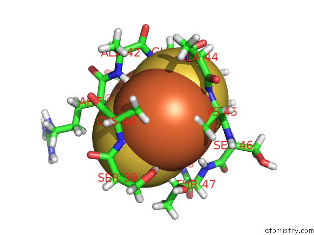
Mono view
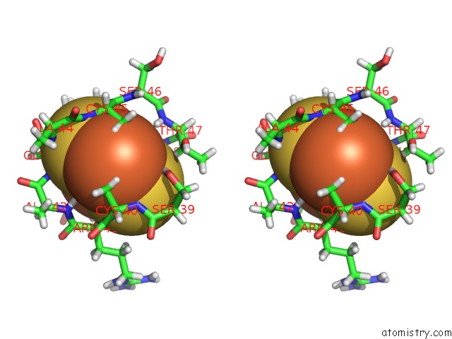
Stereo pair view
Mono view
Stereo pair view
A full contact list of Iron with other atoms in the Fe binding
site number 1 of Crystal Structure Analysis of Cyanidioschyzon Melorae Ferredoxin D58N Mutant within 5.0Å range:
|
Iron binding site 2 out of 2 in 3wcq
Go back to
Iron binding site 2 out
of 2 in the Crystal Structure Analysis of Cyanidioschyzon Melorae Ferredoxin D58N Mutant
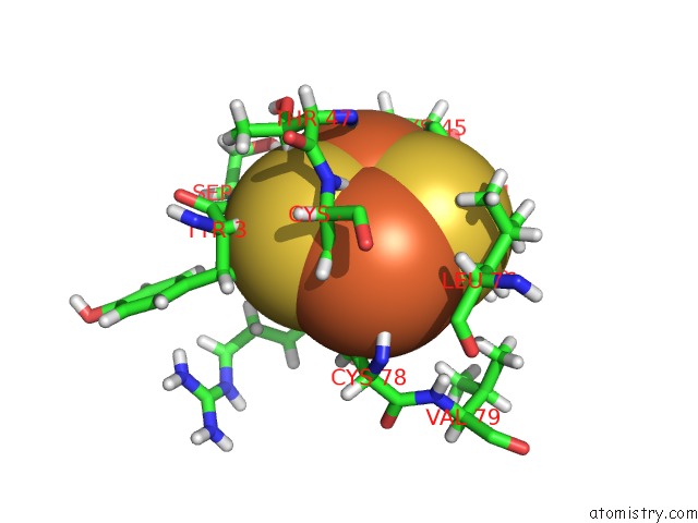
Mono view
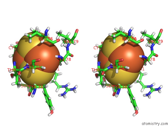
Stereo pair view
Mono view
Stereo pair view
A full contact list of Iron with other atoms in the Fe binding
site number 2 of Crystal Structure Analysis of Cyanidioschyzon Melorae Ferredoxin D58N Mutant within 5.0Å range:
|
Reference:
Y.Ueno,
T.Matsumoto,
A.Yamano,
T.Imai,
Y.Morimoto.
Increasing the Electron-Transfer Ability of Cyanidioschyzon Merolae Ferredoxin By A One-Point Mutation - A High Resolution and Fe-Sad Phasing Crystal Structure Analysis of the ASP58ASN Mutant Biochem.Biophys.Res.Commun. V. 436 736 2013.
ISSN: ISSN 0006-291X
PubMed: 23792094
DOI: 10.1016/J.BBRC.2013.06.029
Page generated: Sun Aug 4 22:35:08 2024
ISSN: ISSN 0006-291X
PubMed: 23792094
DOI: 10.1016/J.BBRC.2013.06.029
Last articles
Zn in 9J0NZn in 9J0O
Zn in 9J0P
Zn in 9FJX
Zn in 9EKB
Zn in 9C0F
Zn in 9CAH
Zn in 9CH0
Zn in 9CH3
Zn in 9CH1