Iron »
PDB 3vtj-3wg7 »
3wec »
Iron in PDB 3wec: Structure of P450 Raua (CYP1050A1) Complexed with A Biosynthetic Intermediate of Aurachin Re
Protein crystallography data
The structure of Structure of P450 Raua (CYP1050A1) Complexed with A Biosynthetic Intermediate of Aurachin Re, PDB code: 3wec
was solved by
Y.Yasutake,
W.Kitagawa,
T.Tamura,
with X-Ray Crystallography technique. A brief refinement statistics is given in the table below:
| Resolution Low / High (Å) | 50.00 / 2.19 |
| Space group | P 1 21 1 |
| Cell size a, b, c (Å), α, β, γ (°) | 41.460, 100.040, 52.290, 90.00, 108.72, 90.00 |
| R / Rfree (%) | 21.4 / 26.3 |
Iron Binding Sites:
The binding sites of Iron atom in the Structure of P450 Raua (CYP1050A1) Complexed with A Biosynthetic Intermediate of Aurachin Re
(pdb code 3wec). This binding sites where shown within
5.0 Angstroms radius around Iron atom.
In total only one binding site of Iron was determined in the Structure of P450 Raua (CYP1050A1) Complexed with A Biosynthetic Intermediate of Aurachin Re, PDB code: 3wec:
In total only one binding site of Iron was determined in the Structure of P450 Raua (CYP1050A1) Complexed with A Biosynthetic Intermediate of Aurachin Re, PDB code: 3wec:
Iron binding site 1 out of 1 in 3wec
Go back to
Iron binding site 1 out
of 1 in the Structure of P450 Raua (CYP1050A1) Complexed with A Biosynthetic Intermediate of Aurachin Re
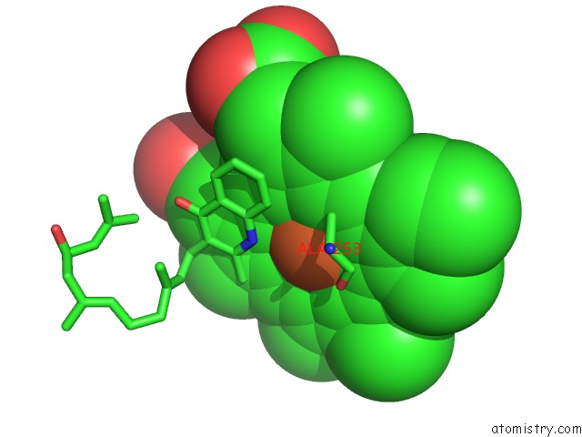
Mono view
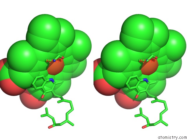
Stereo pair view
Mono view
Stereo pair view
A full contact list of Iron with other atoms in the Fe binding
site number 1 of Structure of P450 Raua (CYP1050A1) Complexed with A Biosynthetic Intermediate of Aurachin Re within 5.0Å range:
|
Reference:
Y.Yasutake,
W.Kitagawa,
M.Hata,
T.Nishioka,
T.Ozaki,
M.Nishiyama,
T.Kuzuyama,
T.Tamura.
Structure of the Quinoline N-Hydroxylating Cytochrome P450 Raua, An Essential Enzyme That Confers Antibiotic Activity on Aurachin Alkaloids Febs Lett. V. 588 105 2014.
ISSN: ISSN 0014-5793
PubMed: 24269679
DOI: 10.1016/J.FEBSLET.2013.11.016
Page generated: Sun Aug 4 22:35:50 2024
ISSN: ISSN 0014-5793
PubMed: 24269679
DOI: 10.1016/J.FEBSLET.2013.11.016
Last articles
Cl in 8BU0Cl in 8BTI
Cl in 8BTE
Cl in 8BTC
Cl in 8BST
Cl in 8BT7
Cl in 8BTA
Cl in 8BT2
Cl in 8BSG
Cl in 8BT1