Iron »
PDB 4b31-4bmp »
4b7g »
Iron in PDB 4b7g: Structure of A Bacterial Catalase
Enzymatic activity of Structure of A Bacterial Catalase
All present enzymatic activity of Structure of A Bacterial Catalase:
1.11.1.6;
1.11.1.6;
Protein crystallography data
The structure of Structure of A Bacterial Catalase, PDB code: 4b7g
was solved by
A.Gumiero,
M.Walsh,
with X-Ray Crystallography technique. A brief refinement statistics is given in the table below:
| Resolution Low / High (Å) | 48.802 / 1.90 |
| Space group | P 63 |
| Cell size a, b, c (Å), α, β, γ (°) | 151.751, 151.751, 157.682, 90.00, 90.00, 120.00 |
| R / Rfree (%) | 13.3 / 17.43 |
Other elements in 4b7g:
The structure of Structure of A Bacterial Catalase also contains other interesting chemical elements:
| Chlorine | (Cl) | 2 atoms |
Iron Binding Sites:
The binding sites of Iron atom in the Structure of A Bacterial Catalase
(pdb code 4b7g). This binding sites where shown within
5.0 Angstroms radius around Iron atom.
In total 4 binding sites of Iron where determined in the Structure of A Bacterial Catalase, PDB code: 4b7g:
Jump to Iron binding site number: 1; 2; 3; 4;
In total 4 binding sites of Iron where determined in the Structure of A Bacterial Catalase, PDB code: 4b7g:
Jump to Iron binding site number: 1; 2; 3; 4;
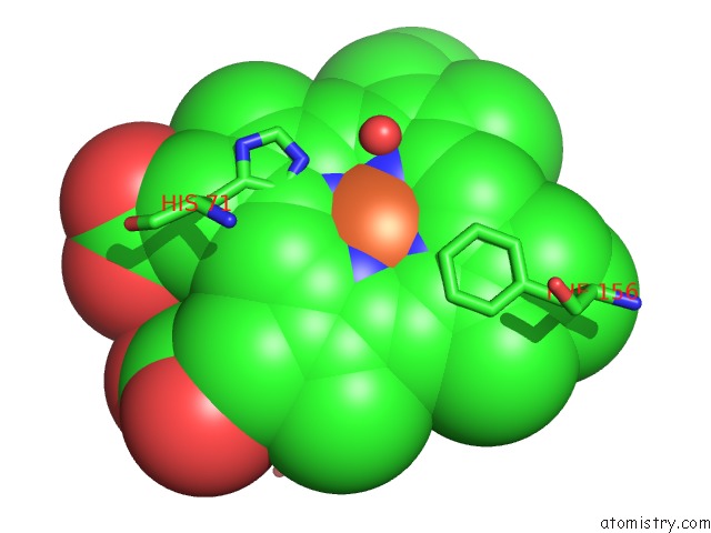
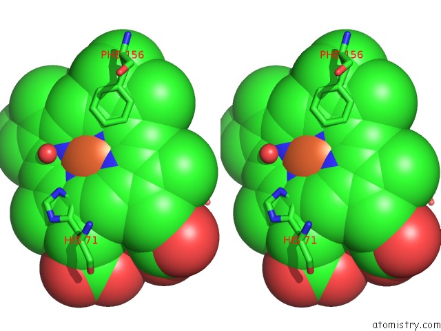
Iron binding site 1 out of 4 in 4b7g
Go back to
Iron binding site 1 out
of 4 in the Structure of A Bacterial Catalase
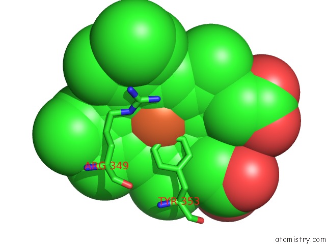
Mono view
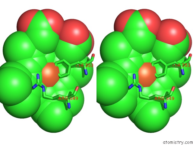
Stereo pair view
Mono view
Stereo pair view
A full contact list of Iron with other atoms in the Fe binding
site number 1 of Structure of A Bacterial Catalase within 5.0Å range:
|
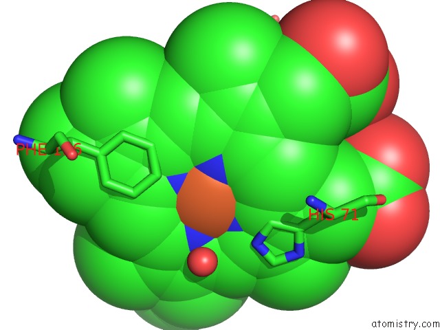
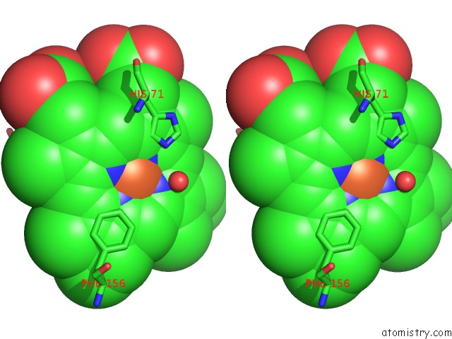
Iron binding site 2 out of 4 in 4b7g
Go back to
Iron binding site 2 out
of 4 in the Structure of A Bacterial Catalase
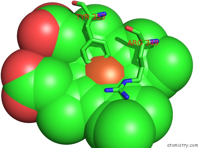
Mono view
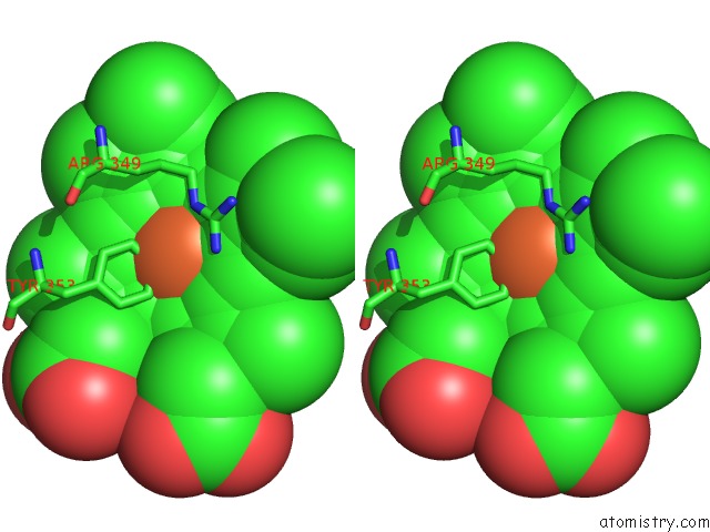
Stereo pair view
Mono view
Stereo pair view
A full contact list of Iron with other atoms in the Fe binding
site number 2 of Structure of A Bacterial Catalase within 5.0Å range:
|
Iron binding site 3 out of 4 in 4b7g
Go back to
Iron binding site 3 out
of 4 in the Structure of A Bacterial Catalase
Mono view
Stereo pair view
Mono view
Stereo pair view
A full contact list of Iron with other atoms in the Fe binding
site number 3 of Structure of A Bacterial Catalase within 5.0Å range:
|
Iron binding site 4 out of 4 in 4b7g
Go back to
Iron binding site 4 out
of 4 in the Structure of A Bacterial Catalase
Mono view
Stereo pair view
Mono view
Stereo pair view
A full contact list of Iron with other atoms in the Fe binding
site number 4 of Structure of A Bacterial Catalase within 5.0Å range:
|
Reference:
M.Candelaresi,
A.Gumiero,
K.Adamczyk,
K.Robb,
C.Bellota-Anton,
V.Sangal,
J.Munnoch,
G.M.Greetham,
M.Towrie,
P.A.Hoskisson,
A.W.Parker,
N.P.Tucker,
M.A.Walsh,
N.T.Hunt.
A Structural and Dynamic Investigation of the Inhibition of Catalase By Nitric Oxide. Org.Biomol.Chem. V. 11 7778 2013.
ISSN: ISSN 1477-0520
PubMed: 24121528
DOI: 10.1039/C3OB41977K
Page generated: Sun Aug 4 23:56:42 2024
ISSN: ISSN 1477-0520
PubMed: 24121528
DOI: 10.1039/C3OB41977K
Last articles
Zn in 9MJ5Zn in 9HNW
Zn in 9G0L
Zn in 9FNE
Zn in 9DZN
Zn in 9E0I
Zn in 9D32
Zn in 9DAK
Zn in 8ZXC
Zn in 8ZUF