Iron »
PDB 4b31-4bmp »
4bk8 »
Iron in PDB 4bk8: Superoxide Reductase (Neelaredoxin) From Ignicoccus Hospitalis
Protein crystallography data
The structure of Superoxide Reductase (Neelaredoxin) From Ignicoccus Hospitalis, PDB code: 4bk8
was solved by
C.V.Romao,
P.M.Matias,
F.G.Pinho,
C.M.Sousa,
A.R.Barradas,
A.F.Pinto,
M.Teixeira,
T.M.Bandeiras,
with X-Ray Crystallography technique. A brief refinement statistics is given in the table below:
| Resolution Low / High (Å) | 47.196 / 1.85 |
| Space group | P 64 2 2 |
| Cell size a, b, c (Å), α, β, γ (°) | 108.995, 108.995, 61.430, 90.00, 90.00, 120.00 |
| R / Rfree (%) | 17.77 / 19.95 |
Iron Binding Sites:
The binding sites of Iron atom in the Superoxide Reductase (Neelaredoxin) From Ignicoccus Hospitalis
(pdb code 4bk8). This binding sites where shown within
5.0 Angstroms radius around Iron atom.
In total only one binding site of Iron was determined in the Superoxide Reductase (Neelaredoxin) From Ignicoccus Hospitalis, PDB code: 4bk8:
In total only one binding site of Iron was determined in the Superoxide Reductase (Neelaredoxin) From Ignicoccus Hospitalis, PDB code: 4bk8:
Iron binding site 1 out of 1 in 4bk8
Go back to
Iron binding site 1 out
of 1 in the Superoxide Reductase (Neelaredoxin) From Ignicoccus Hospitalis
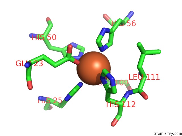
Mono view
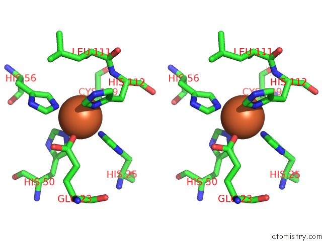
Stereo pair view
Mono view
Stereo pair view
A full contact list of Iron with other atoms in the Fe binding
site number 1 of Superoxide Reductase (Neelaredoxin) From Ignicoccus Hospitalis within 5.0Å range:
|
Reference:
F.G.Pinho,
C.V.Romao,
A.F.Pinto,
P.M.Matias,
M.Teixeira,
T.M.Bandeiras.
Structure of A Natural Sor Mutant To Be Published.
Page generated: Tue Aug 5 09:14:31 2025
Last articles
Fe in 5F0BFe in 5EZW
Fe in 5EYS
Fe in 5EYJ
Fe in 5EXR
Fe in 5EXQ
Fe in 5EXJ
Fe in 5EXI
Fe in 5EX6
Fe in 5EX8