Iron »
PDB 4dig-4egm »
4dtw »
Iron in PDB 4dtw: Cytochrome P450 BM3H-8C8 Mri Sensor Bound to Serotonin
Enzymatic activity of Cytochrome P450 BM3H-8C8 Mri Sensor Bound to Serotonin
All present enzymatic activity of Cytochrome P450 BM3H-8C8 Mri Sensor Bound to Serotonin:
1.14.14.1;
1.14.14.1;
Protein crystallography data
The structure of Cytochrome P450 BM3H-8C8 Mri Sensor Bound to Serotonin, PDB code: 4dtw
was solved by
E.M.Brustad,
V.S.Lelyveld,
C.D.Snow,
N.Crook,
F.M.Martinez,
T.J.Scholl,
A.Jasanoff,
F.H.Arnold,
with X-Ray Crystallography technique. A brief refinement statistics is given in the table below:
| Resolution Low / High (Å) | 38.87 / 1.80 |
| Space group | P 1 21 1 |
| Cell size a, b, c (Å), α, β, γ (°) | 58.606, 146.249, 64.064, 90.00, 97.51, 90.00 |
| R / Rfree (%) | 19.2 / 25.4 |
Other elements in 4dtw:
The structure of Cytochrome P450 BM3H-8C8 Mri Sensor Bound to Serotonin also contains other interesting chemical elements:
| Magnesium | (Mg) | 1 atom |
Iron Binding Sites:
The binding sites of Iron atom in the Cytochrome P450 BM3H-8C8 Mri Sensor Bound to Serotonin
(pdb code 4dtw). This binding sites where shown within
5.0 Angstroms radius around Iron atom.
In total 2 binding sites of Iron where determined in the Cytochrome P450 BM3H-8C8 Mri Sensor Bound to Serotonin, PDB code: 4dtw:
Jump to Iron binding site number: 1; 2;
In total 2 binding sites of Iron where determined in the Cytochrome P450 BM3H-8C8 Mri Sensor Bound to Serotonin, PDB code: 4dtw:
Jump to Iron binding site number: 1; 2;
Iron binding site 1 out of 2 in 4dtw
Go back to
Iron binding site 1 out
of 2 in the Cytochrome P450 BM3H-8C8 Mri Sensor Bound to Serotonin
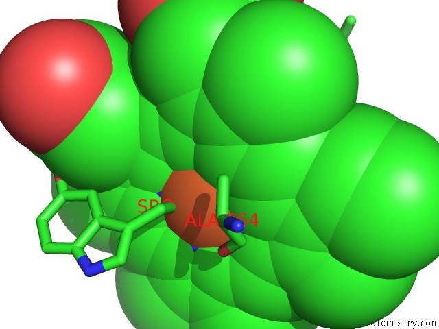
Mono view
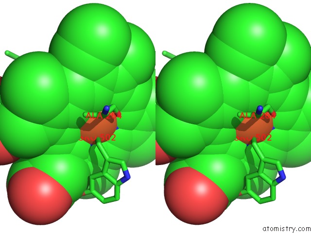
Stereo pair view
Mono view
Stereo pair view
A full contact list of Iron with other atoms in the Fe binding
site number 1 of Cytochrome P450 BM3H-8C8 Mri Sensor Bound to Serotonin within 5.0Å range:
|
Iron binding site 2 out of 2 in 4dtw
Go back to
Iron binding site 2 out
of 2 in the Cytochrome P450 BM3H-8C8 Mri Sensor Bound to Serotonin
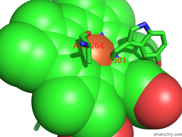
Mono view
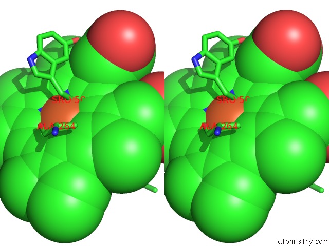
Stereo pair view
Mono view
Stereo pair view
A full contact list of Iron with other atoms in the Fe binding
site number 2 of Cytochrome P450 BM3H-8C8 Mri Sensor Bound to Serotonin within 5.0Å range:
|
Reference:
E.M.Brustad,
V.S.Lelyveld,
C.D.Snow,
N.Crook,
S.T.Jung,
F.M.Martinez,
T.J.Scholl,
A.Jasanoff,
F.H.Arnold.
Structure-Guided Directed Evolution of Highly Selective P450-Based Magnetic Resonance Imaging Sensors For Dopamine and Serotonin. J.Mol.Biol. V. 422 245 2012.
ISSN: ISSN 0022-2836
PubMed: 22659321
DOI: 10.1016/J.JMB.2012.05.029
Page generated: Tue Aug 5 09:54:42 2025
ISSN: ISSN 0022-2836
PubMed: 22659321
DOI: 10.1016/J.JMB.2012.05.029
Last articles
Fe in 4NVJFe in 4NVK
Fe in 4NVI
Fe in 4NVH
Fe in 4NVG
Fe in 4NVF
Fe in 4NVE
Fe in 4NVD
Fe in 4NVC
Fe in 4NSE