Iron »
PDB 4egn-4f2n »
4erm »
Iron in PDB 4erm: Crystal Structure of the Datp Inhibited E. Coli Class Ia Ribonucleotide Reductase Complex at 4 Angstroms Resolution
Enzymatic activity of Crystal Structure of the Datp Inhibited E. Coli Class Ia Ribonucleotide Reductase Complex at 4 Angstroms Resolution
All present enzymatic activity of Crystal Structure of the Datp Inhibited E. Coli Class Ia Ribonucleotide Reductase Complex at 4 Angstroms Resolution:
1.17.4.1;
1.17.4.1;
Protein crystallography data
The structure of Crystal Structure of the Datp Inhibited E. Coli Class Ia Ribonucleotide Reductase Complex at 4 Angstroms Resolution, PDB code: 4erm
was solved by
C.M.Zimanyi,
C.L.Drennan,
with X-Ray Crystallography technique. A brief refinement statistics is given in the table below:
| Resolution Low / High (Å) | 30.00 / 3.95 |
| Space group | C 1 2 1 |
| Cell size a, b, c (Å), α, β, γ (°) | 280.471, 155.744, 166.919, 90.00, 119.07, 90.00 |
| R / Rfree (%) | 25.9 / 28.4 |
Other elements in 4erm:
The structure of Crystal Structure of the Datp Inhibited E. Coli Class Ia Ribonucleotide Reductase Complex at 4 Angstroms Resolution also contains other interesting chemical elements:
| Magnesium | (Mg) | 4 atoms |
Iron Binding Sites:
The binding sites of Iron atom in the Crystal Structure of the Datp Inhibited E. Coli Class Ia Ribonucleotide Reductase Complex at 4 Angstroms Resolution
(pdb code 4erm). This binding sites where shown within
5.0 Angstroms radius around Iron atom.
In total 8 binding sites of Iron where determined in the Crystal Structure of the Datp Inhibited E. Coli Class Ia Ribonucleotide Reductase Complex at 4 Angstroms Resolution, PDB code: 4erm:
Jump to Iron binding site number: 1; 2; 3; 4; 5; 6; 7; 8;
In total 8 binding sites of Iron where determined in the Crystal Structure of the Datp Inhibited E. Coli Class Ia Ribonucleotide Reductase Complex at 4 Angstroms Resolution, PDB code: 4erm:
Jump to Iron binding site number: 1; 2; 3; 4; 5; 6; 7; 8;
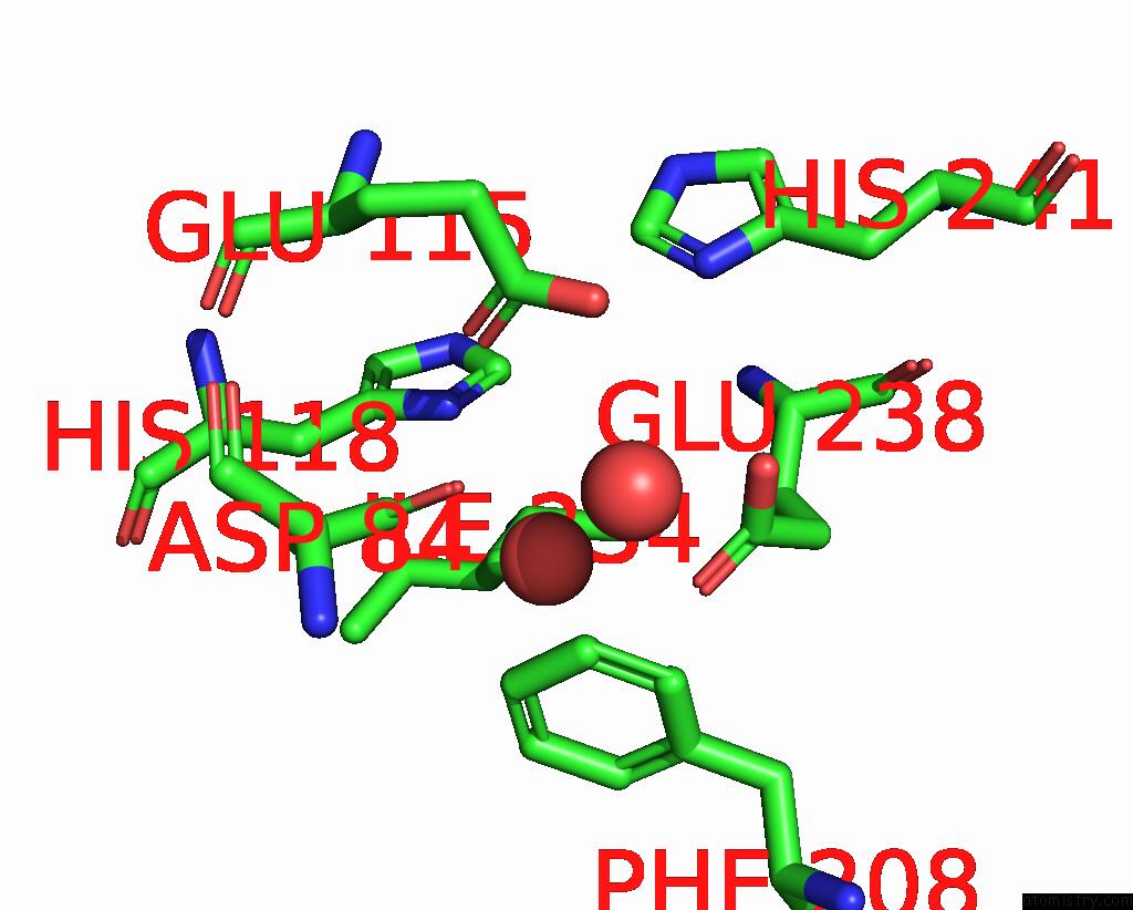
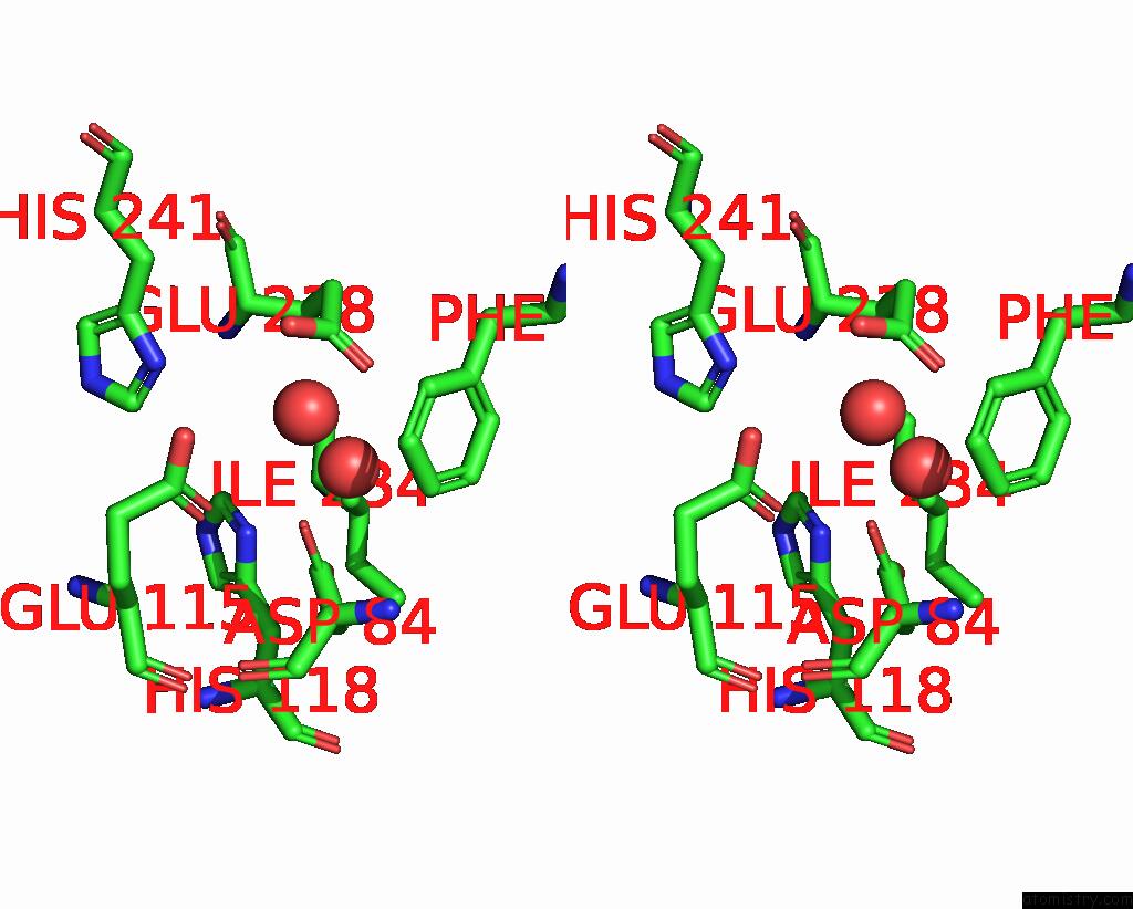
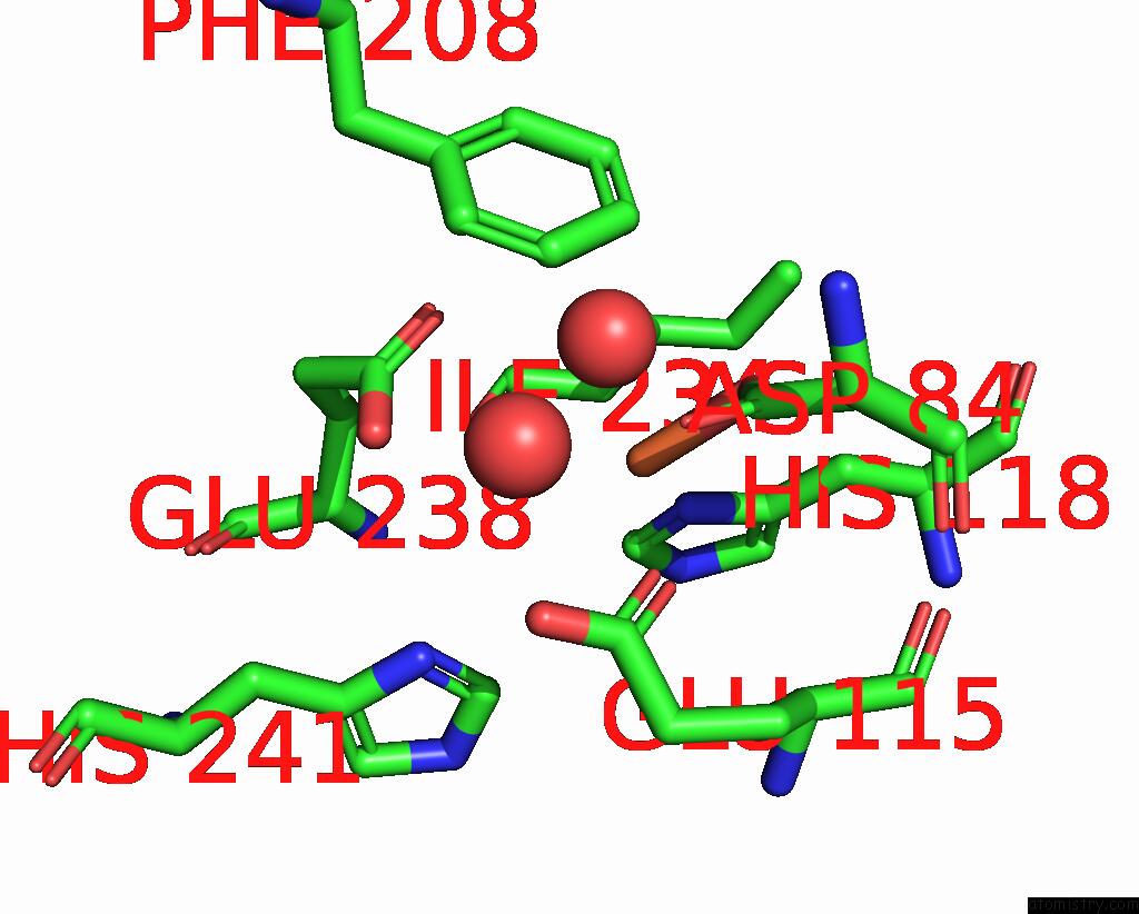
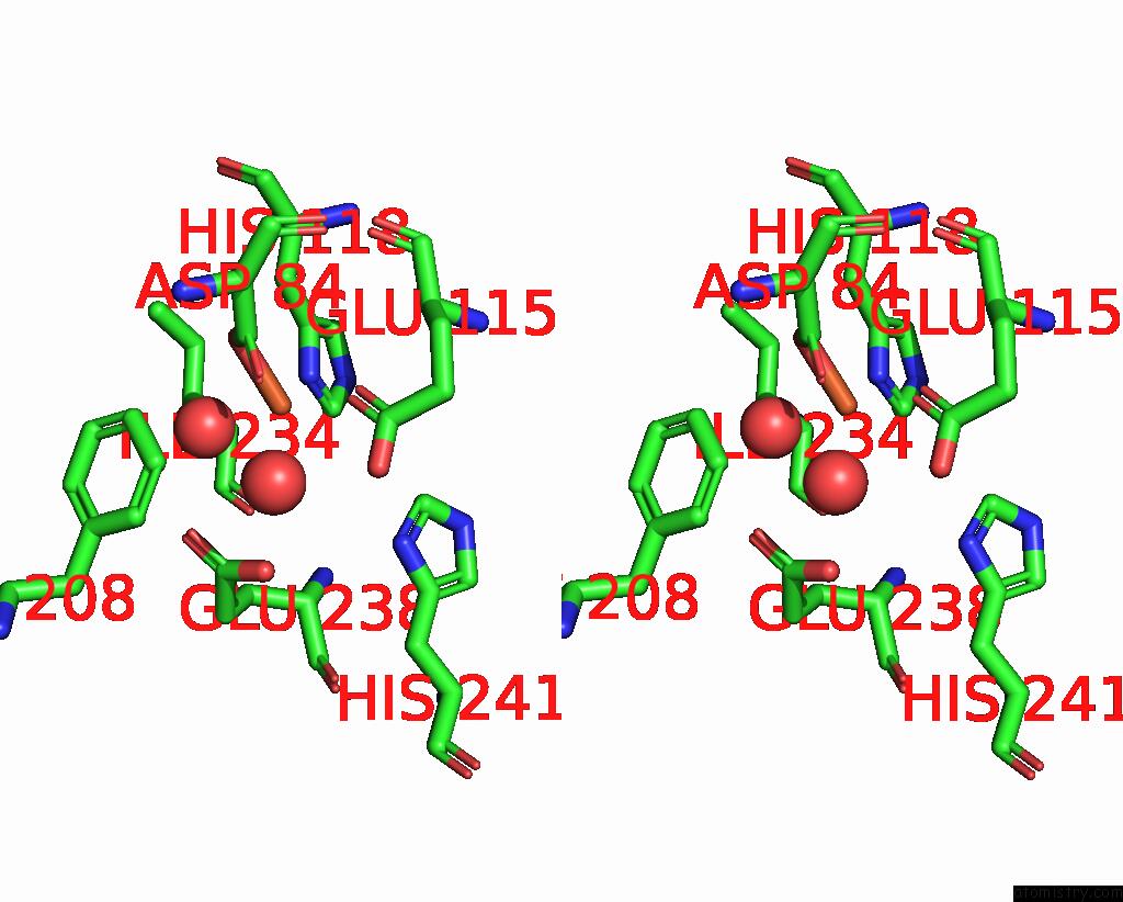
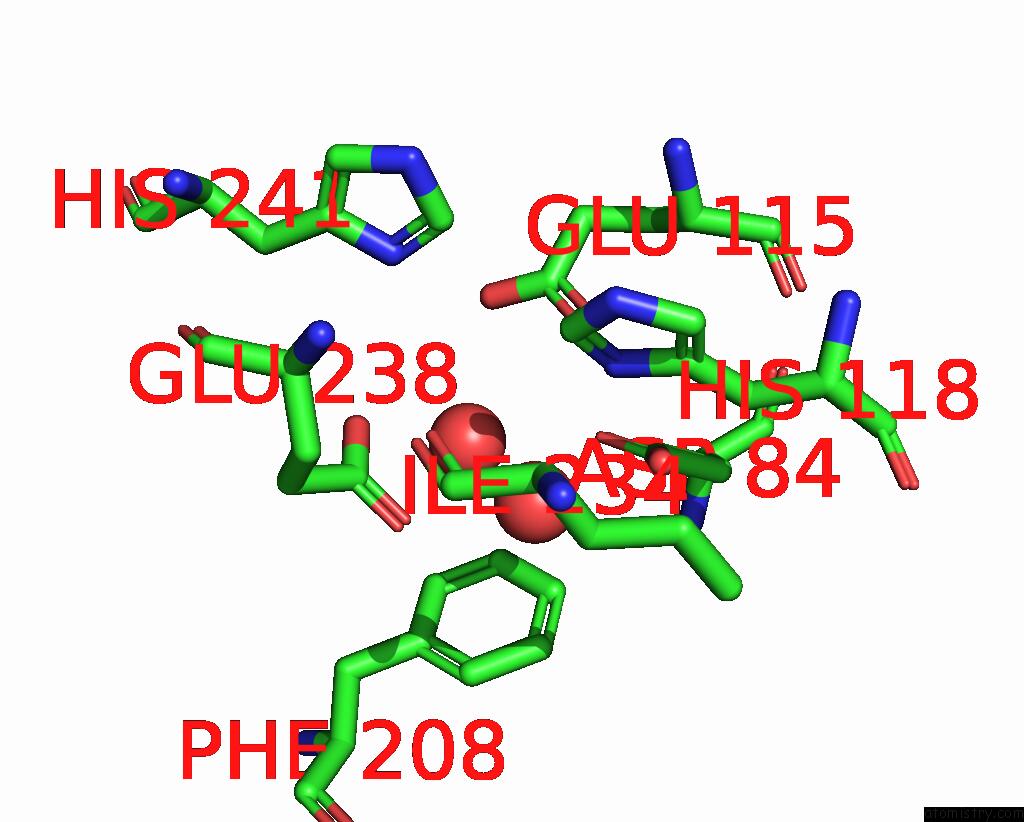
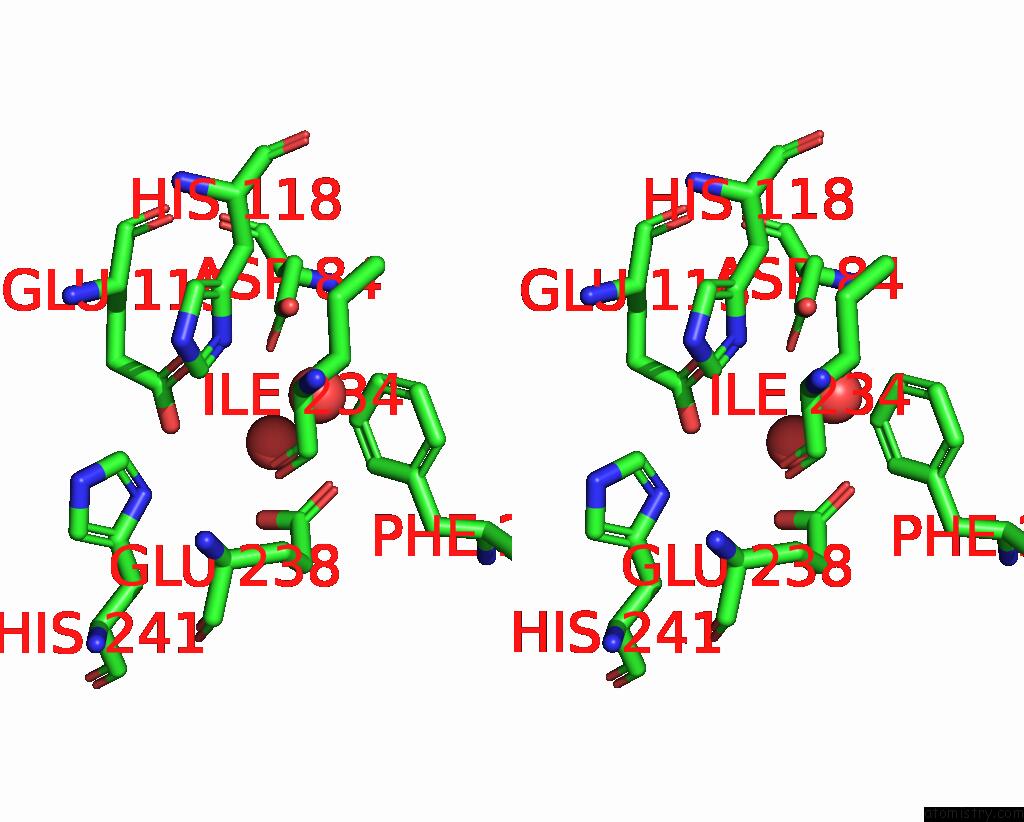
Iron binding site 1 out of 8 in 4erm
Go back to
Iron binding site 1 out
of 8 in the Crystal Structure of the Datp Inhibited E. Coli Class Ia Ribonucleotide Reductase Complex at 4 Angstroms Resolution
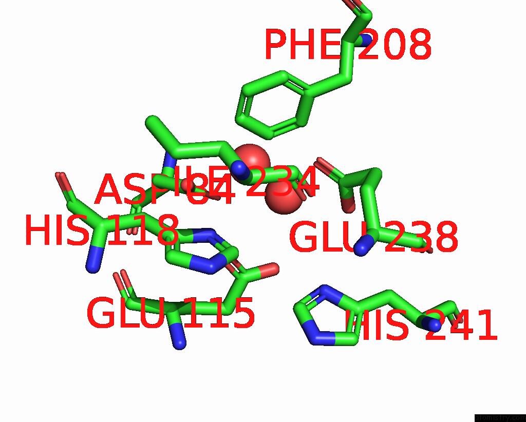
Mono view
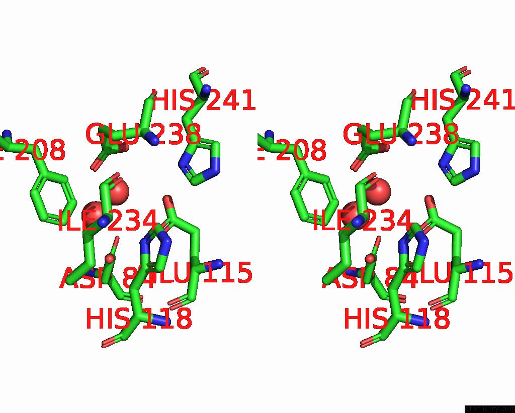
Stereo pair view
Mono view
Stereo pair view
A full contact list of Iron with other atoms in the Fe binding
site number 1 of Crystal Structure of the Datp Inhibited E. Coli Class Ia Ribonucleotide Reductase Complex at 4 Angstroms Resolution within 5.0Å range:
|
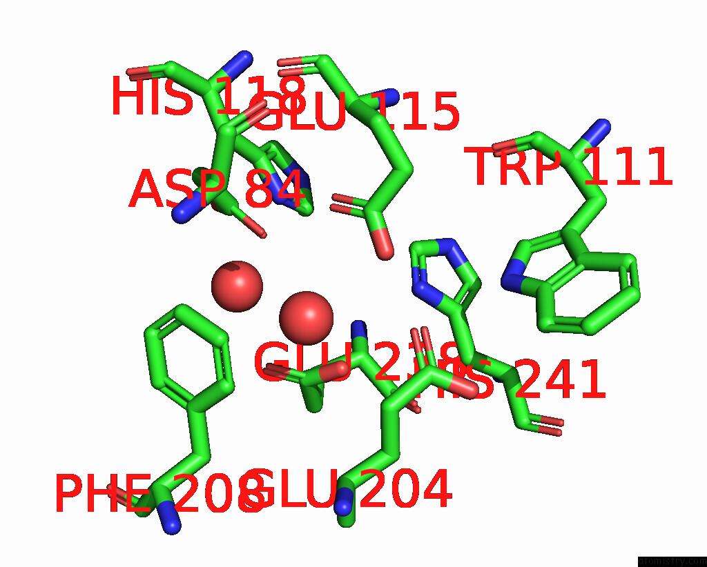
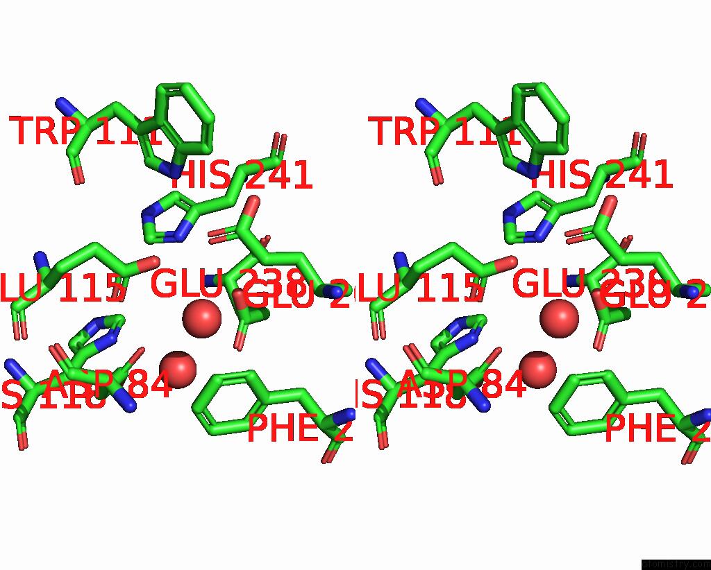
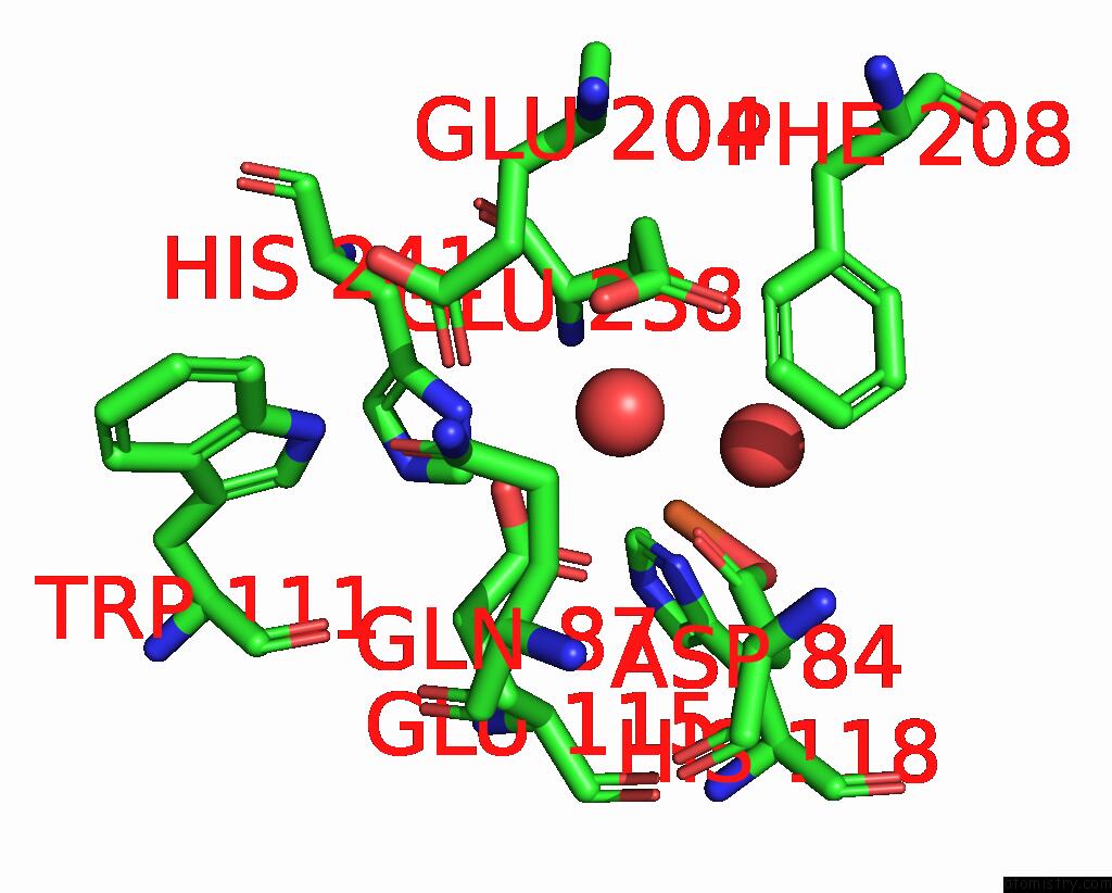
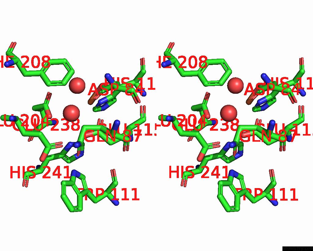
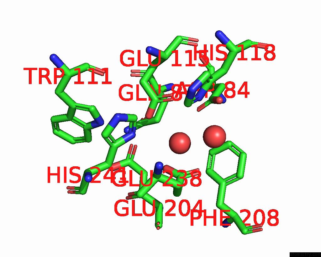
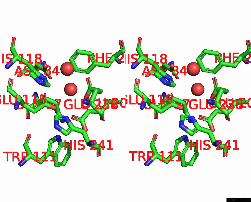
Iron binding site 2 out of 8 in 4erm
Go back to
Iron binding site 2 out
of 8 in the Crystal Structure of the Datp Inhibited E. Coli Class Ia Ribonucleotide Reductase Complex at 4 Angstroms Resolution
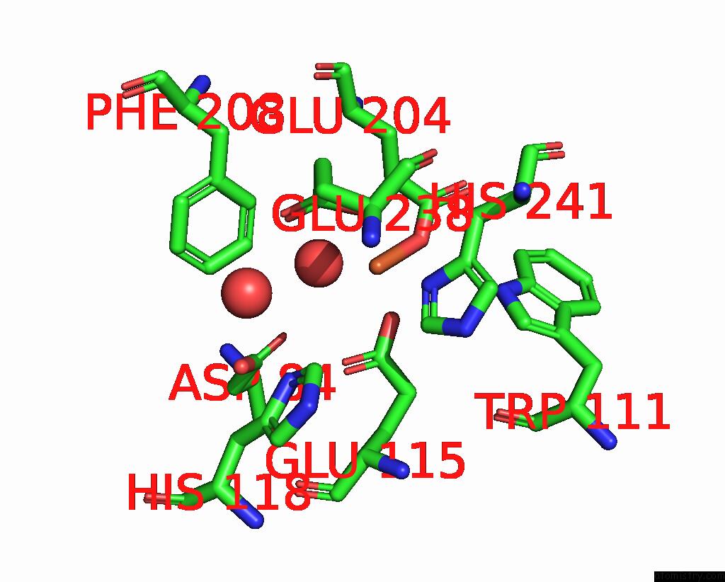
Mono view
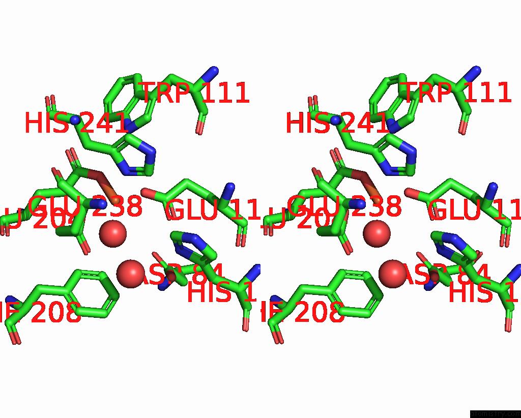
Stereo pair view
Mono view
Stereo pair view
A full contact list of Iron with other atoms in the Fe binding
site number 2 of Crystal Structure of the Datp Inhibited E. Coli Class Ia Ribonucleotide Reductase Complex at 4 Angstroms Resolution within 5.0Å range:
|
Iron binding site 3 out of 8 in 4erm
Go back to
Iron binding site 3 out
of 8 in the Crystal Structure of the Datp Inhibited E. Coli Class Ia Ribonucleotide Reductase Complex at 4 Angstroms Resolution
Mono view
Stereo pair view
Mono view
Stereo pair view
A full contact list of Iron with other atoms in the Fe binding
site number 3 of Crystal Structure of the Datp Inhibited E. Coli Class Ia Ribonucleotide Reductase Complex at 4 Angstroms Resolution within 5.0Å range:
|
Iron binding site 4 out of 8 in 4erm
Go back to
Iron binding site 4 out
of 8 in the Crystal Structure of the Datp Inhibited E. Coli Class Ia Ribonucleotide Reductase Complex at 4 Angstroms Resolution
Mono view
Stereo pair view
Mono view
Stereo pair view
A full contact list of Iron with other atoms in the Fe binding
site number 4 of Crystal Structure of the Datp Inhibited E. Coli Class Ia Ribonucleotide Reductase Complex at 4 Angstroms Resolution within 5.0Å range:
|
Iron binding site 5 out of 8 in 4erm
Go back to
Iron binding site 5 out
of 8 in the Crystal Structure of the Datp Inhibited E. Coli Class Ia Ribonucleotide Reductase Complex at 4 Angstroms Resolution
Mono view
Stereo pair view
Mono view
Stereo pair view
A full contact list of Iron with other atoms in the Fe binding
site number 5 of Crystal Structure of the Datp Inhibited E. Coli Class Ia Ribonucleotide Reductase Complex at 4 Angstroms Resolution within 5.0Å range:
|
Iron binding site 6 out of 8 in 4erm
Go back to
Iron binding site 6 out
of 8 in the Crystal Structure of the Datp Inhibited E. Coli Class Ia Ribonucleotide Reductase Complex at 4 Angstroms Resolution
Mono view
Stereo pair view
Mono view
Stereo pair view
A full contact list of Iron with other atoms in the Fe binding
site number 6 of Crystal Structure of the Datp Inhibited E. Coli Class Ia Ribonucleotide Reductase Complex at 4 Angstroms Resolution within 5.0Å range:
|
Iron binding site 7 out of 8 in 4erm
Go back to
Iron binding site 7 out
of 8 in the Crystal Structure of the Datp Inhibited E. Coli Class Ia Ribonucleotide Reductase Complex at 4 Angstroms Resolution
Mono view
Stereo pair view
Mono view
Stereo pair view
A full contact list of Iron with other atoms in the Fe binding
site number 7 of Crystal Structure of the Datp Inhibited E. Coli Class Ia Ribonucleotide Reductase Complex at 4 Angstroms Resolution within 5.0Å range:
|
Iron binding site 8 out of 8 in 4erm
Go back to
Iron binding site 8 out
of 8 in the Crystal Structure of the Datp Inhibited E. Coli Class Ia Ribonucleotide Reductase Complex at 4 Angstroms Resolution
Mono view
Stereo pair view
Mono view
Stereo pair view
A full contact list of Iron with other atoms in the Fe binding
site number 8 of Crystal Structure of the Datp Inhibited E. Coli Class Ia Ribonucleotide Reductase Complex at 4 Angstroms Resolution within 5.0Å range:
|
Reference:
C.M.Zimanyi,
N.Ando,
E.J.Brignole,
F.J.Asturias,
J.Stubbe,
C.L.Drennan.
Tangled Up in Knots: Structures of Inactivated Forms of E. Coli Class Ia Ribonucleotide Reductase. Structure V. 20 1374 2012.
ISSN: ISSN 0969-2126
PubMed: 22727814
DOI: 10.1016/J.STR.2012.05.009
Page generated: Tue Aug 5 10:10:20 2025
ISSN: ISSN 0969-2126
PubMed: 22727814
DOI: 10.1016/J.STR.2012.05.009
Last articles
Fe in 4Q5OFe in 4Q1O
Fe in 4Q17
Fe in 4Q0T
Fe in 4PV1
Fe in 4PWA
Fe in 4PXH
Fe in 4PL2
Fe in 4PWV
Fe in 4PL1