Iron »
PDB 4fh7-4g38 »
4fjp »
Iron in PDB 4fjp: Crystal Structure of C-Lobe of Bovine Lactoferrin Complexed with Naproxen at 1.68 A Resolution
Protein crystallography data
The structure of Crystal Structure of C-Lobe of Bovine Lactoferrin Complexed with Naproxen at 1.68 A Resolution, PDB code: 4fjp
was solved by
P.K.Shukla,
L.Gautam,
M.Sinha,
P.Kaur,
S.Sharma,
T.P.Singh,
with X-Ray Crystallography technique. A brief refinement statistics is given in the table below:
| Resolution Low / High (Å) | 62.45 / 1.68 |
| Space group | P 1 21 1 |
| Cell size a, b, c (Å), α, β, γ (°) | 62.277, 49.945, 65.301, 90.00, 107.00, 90.00 |
| R / Rfree (%) | 16.3 / 20.8 |
Other elements in 4fjp:
The structure of Crystal Structure of C-Lobe of Bovine Lactoferrin Complexed with Naproxen at 1.68 A Resolution also contains other interesting chemical elements:
| Zinc | (Zn) | 2 atoms |
Iron Binding Sites:
The binding sites of Iron atom in the Crystal Structure of C-Lobe of Bovine Lactoferrin Complexed with Naproxen at 1.68 A Resolution
(pdb code 4fjp). This binding sites where shown within
5.0 Angstroms radius around Iron atom.
In total only one binding site of Iron was determined in the Crystal Structure of C-Lobe of Bovine Lactoferrin Complexed with Naproxen at 1.68 A Resolution, PDB code: 4fjp:
In total only one binding site of Iron was determined in the Crystal Structure of C-Lobe of Bovine Lactoferrin Complexed with Naproxen at 1.68 A Resolution, PDB code: 4fjp:
Iron binding site 1 out of 1 in 4fjp
Go back to
Iron binding site 1 out
of 1 in the Crystal Structure of C-Lobe of Bovine Lactoferrin Complexed with Naproxen at 1.68 A Resolution
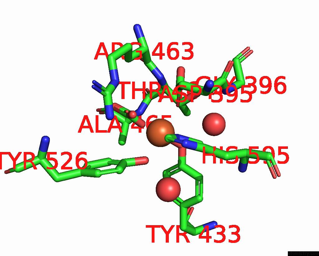
Mono view
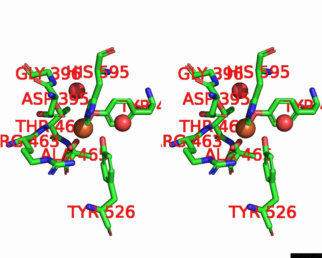
Stereo pair view
Mono view
Stereo pair view
A full contact list of Iron with other atoms in the Fe binding
site number 1 of Crystal Structure of C-Lobe of Bovine Lactoferrin Complexed with Naproxen at 1.68 A Resolution within 5.0Å range:
|
Reference:
P.K.Shukla,
L.Gautam,
M.Sinha,
P.Kaur,
S.Sharma,
T.P.Singh.
Crystal Structure of C-Lobe of Bovine Lactoferrin Complexed with Naproxen at 1.68 A Resolution To Be Published.
Page generated: Tue Aug 5 10:27:50 2025
Last articles
Fe in 4O4TFe in 4O1Z
Fe in 4O35
Fe in 4O2G
Fe in 4O1Q
Fe in 4NZI
Fe in 4O1T
Fe in 4NZ2
Fe in 4NY4
Fe in 4NXC