Iron »
PDB 4ryx-4toe »
4s36 »
Iron in PDB 4s36: Crystal Structure of the C-Terminal Domain of R2 Pyocin Membrane- Piercing Spike
Protein crystallography data
The structure of Crystal Structure of the C-Terminal Domain of R2 Pyocin Membrane- Piercing Spike, PDB code: 4s36
was solved by
C.B.Browning,
P.G.Leiman,
M.M.Shneider,
with X-Ray Crystallography technique. A brief refinement statistics is given in the table below:
| Resolution Low / High (Å) | 40.02 / 1.46 |
| Space group | P 63 2 2 |
| Cell size a, b, c (Å), α, β, γ (°) | 46.205, 46.205, 144.954, 90.00, 90.00, 120.00 |
| R / Rfree (%) | 14.9 / 18.8 |
Iron Binding Sites:
The binding sites of Iron atom in the Crystal Structure of the C-Terminal Domain of R2 Pyocin Membrane- Piercing Spike
(pdb code 4s36). This binding sites where shown within
5.0 Angstroms radius around Iron atom.
In total only one binding site of Iron was determined in the Crystal Structure of the C-Terminal Domain of R2 Pyocin Membrane- Piercing Spike, PDB code: 4s36:
In total only one binding site of Iron was determined in the Crystal Structure of the C-Terminal Domain of R2 Pyocin Membrane- Piercing Spike, PDB code: 4s36:
Iron binding site 1 out of 1 in 4s36
Go back to
Iron binding site 1 out
of 1 in the Crystal Structure of the C-Terminal Domain of R2 Pyocin Membrane- Piercing Spike
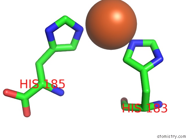
Mono view
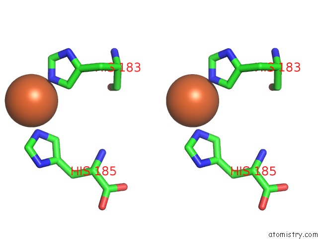
Stereo pair view
Mono view
Stereo pair view
A full contact list of Iron with other atoms in the Fe binding
site number 1 of Crystal Structure of the C-Terminal Domain of R2 Pyocin Membrane- Piercing Spike within 5.0Å range:
|
Reference:
C.B.Browning,
P.G.Leiman,
M.M.Shneider.
Crystal Structure of the C-Terminal Domain of R2 Pyocin Membrane-Piercing Spike To Be Published.
Page generated: Tue Aug 5 14:31:44 2025
Last articles
Fe in 6CYYFe in 6CYJ
Fe in 6CYP
Fe in 6CYS
Fe in 6CXV
Fe in 6CXU
Fe in 6CSB
Fe in 6CWW
Fe in 6CVC
Fe in 6CUN