Iron »
PDB 4z6q-4zn3 »
4zhk »
Iron in PDB 4zhk: Crystal Structure of RPE65 in Complex with Mb-002
Enzymatic activity of Crystal Structure of RPE65 in Complex with Mb-002
All present enzymatic activity of Crystal Structure of RPE65 in Complex with Mb-002:
3.1.1.64;
3.1.1.64;
Protein crystallography data
The structure of Crystal Structure of RPE65 in Complex with Mb-002, PDB code: 4zhk
was solved by
P.D.Kiser,
K.Palczewski,
with X-Ray Crystallography technique. A brief refinement statistics is given in the table below:
| Resolution Low / High (Å) | 47.85 / 2.09 |
| Space group | P 65 |
| Cell size a, b, c (Å), α, β, γ (°) | 175.668, 175.668, 86.297, 90.00, 90.00, 120.00 |
| R / Rfree (%) | 17.2 / 20.5 |
Other elements in 4zhk:
The structure of Crystal Structure of RPE65 in Complex with Mb-002 also contains other interesting chemical elements:
| Sodium | (Na) | 2 atoms |
Iron Binding Sites:
The binding sites of Iron atom in the Crystal Structure of RPE65 in Complex with Mb-002
(pdb code 4zhk). This binding sites where shown within
5.0 Angstroms radius around Iron atom.
In total 2 binding sites of Iron where determined in the Crystal Structure of RPE65 in Complex with Mb-002, PDB code: 4zhk:
Jump to Iron binding site number: 1; 2;
In total 2 binding sites of Iron where determined in the Crystal Structure of RPE65 in Complex with Mb-002, PDB code: 4zhk:
Jump to Iron binding site number: 1; 2;
Iron binding site 1 out of 2 in 4zhk
Go back to
Iron binding site 1 out
of 2 in the Crystal Structure of RPE65 in Complex with Mb-002
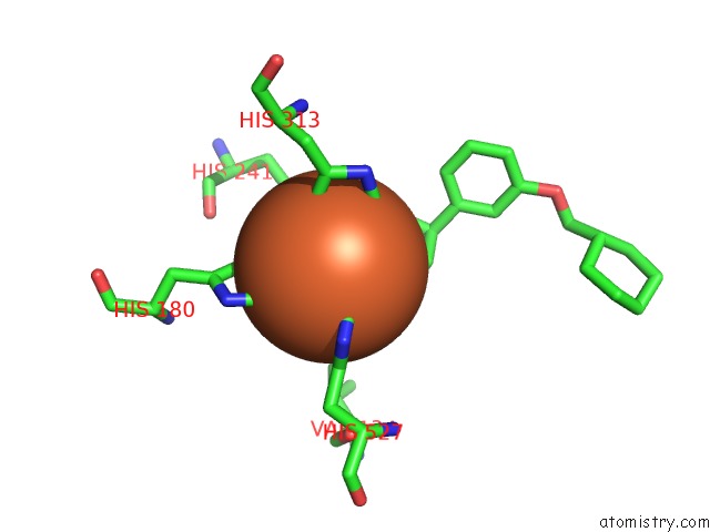
Mono view

Stereo pair view
Mono view
Stereo pair view
A full contact list of Iron with other atoms in the Fe binding
site number 1 of Crystal Structure of RPE65 in Complex with Mb-002 within 5.0Å range:
|
Iron binding site 2 out of 2 in 4zhk
Go back to
Iron binding site 2 out
of 2 in the Crystal Structure of RPE65 in Complex with Mb-002
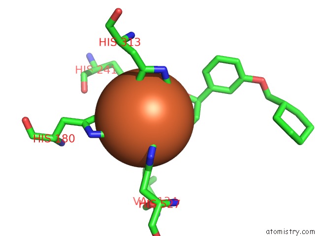
Mono view
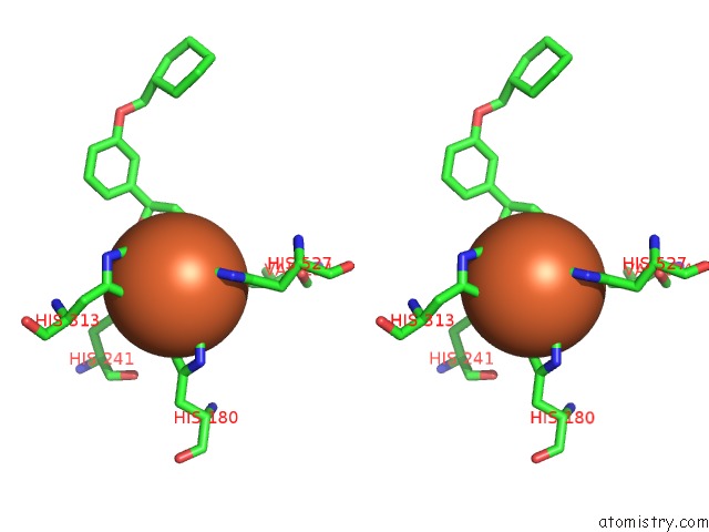
Stereo pair view
Mono view
Stereo pair view
A full contact list of Iron with other atoms in the Fe binding
site number 2 of Crystal Structure of RPE65 in Complex with Mb-002 within 5.0Å range:
|
Reference:
P.D.Kiser,
K.Palczewski.
Crystal Structure of RPE65 in Complex with Mb-002 To Be Published.
Page generated: Tue Aug 5 18:28:39 2025
Last articles
Fe in 6L1BFe in 6L1A
Fe in 6L32
Fe in 6L2J
Fe in 6L1X
Fe in 6KUM
Fe in 6KZT
Fe in 6KV0
Fe in 6KZS
Fe in 6KW7