Iron »
PDB 5g6f-5h8y »
5gn7 »
Iron in PDB 5gn7: Crystal Structure of Alternative Oxidase From Trypanosoma Brucei Brucei Complexed with Cumarin Derivative-17
Protein crystallography data
The structure of Crystal Structure of Alternative Oxidase From Trypanosoma Brucei Brucei Complexed with Cumarin Derivative-17, PDB code: 5gn7
was solved by
E.O.Balogun,
D.K.Inaoka,
T.Shiba,
T.Tsuge,
B.May,
T.Sato,
Y.Kido,
N.Takeshi,
T.Aoki,
T.Honma,
A.Tanaka,
M.Inoue,
S.Matsuoka,
P.A.M.Michels,
Y.Watanabe,
A.L.Moore,
S.Harada,
K.Kita,
with X-Ray Crystallography technique. A brief refinement statistics is given in the table below:
| Resolution Low / High (Å) | 45.54 / 3.20 |
| Space group | C 1 2 1 |
| Cell size a, b, c (Å), α, β, γ (°) | 151.312, 222.726, 63.165, 90.00, 114.70, 90.00 |
| R / Rfree (%) | 19.5 / 26.8 |
Iron Binding Sites:
The binding sites of Iron atom in the Crystal Structure of Alternative Oxidase From Trypanosoma Brucei Brucei Complexed with Cumarin Derivative-17
(pdb code 5gn7). This binding sites where shown within
5.0 Angstroms radius around Iron atom.
In total 8 binding sites of Iron where determined in the Crystal Structure of Alternative Oxidase From Trypanosoma Brucei Brucei Complexed with Cumarin Derivative-17, PDB code: 5gn7:
Jump to Iron binding site number: 1; 2; 3; 4; 5; 6; 7; 8;
In total 8 binding sites of Iron where determined in the Crystal Structure of Alternative Oxidase From Trypanosoma Brucei Brucei Complexed with Cumarin Derivative-17, PDB code: 5gn7:
Jump to Iron binding site number: 1; 2; 3; 4; 5; 6; 7; 8;
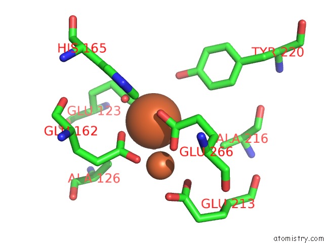
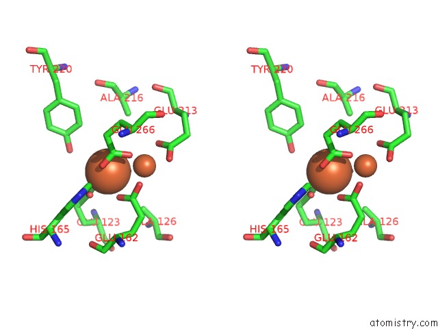
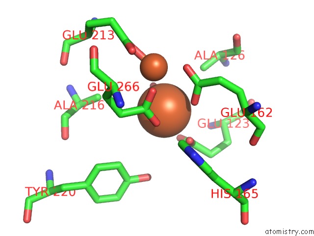
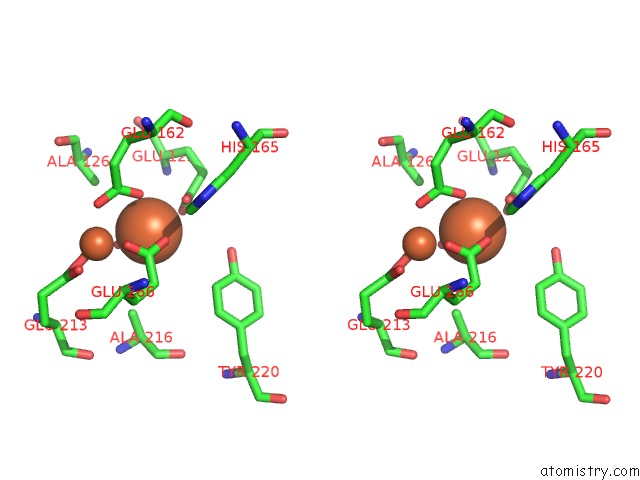
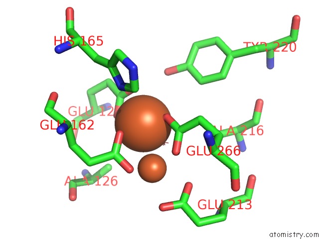
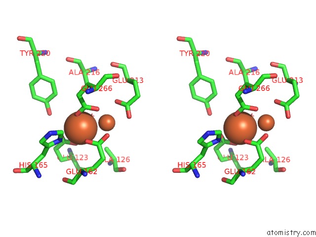
Iron binding site 1 out of 8 in 5gn7
Go back to
Iron binding site 1 out
of 8 in the Crystal Structure of Alternative Oxidase From Trypanosoma Brucei Brucei Complexed with Cumarin Derivative-17
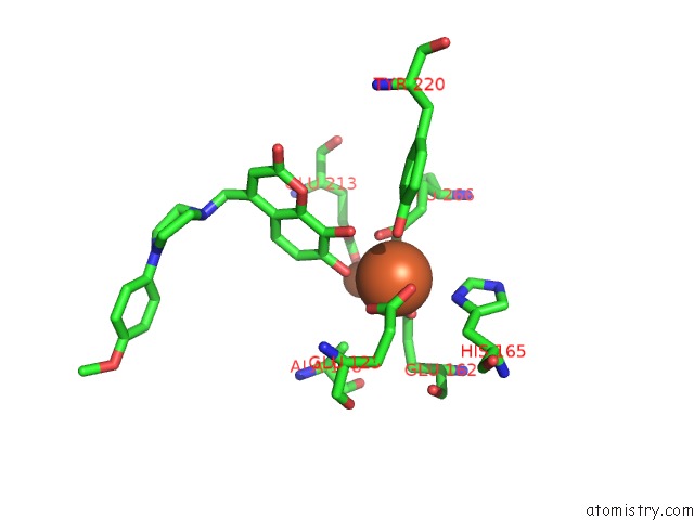
Mono view
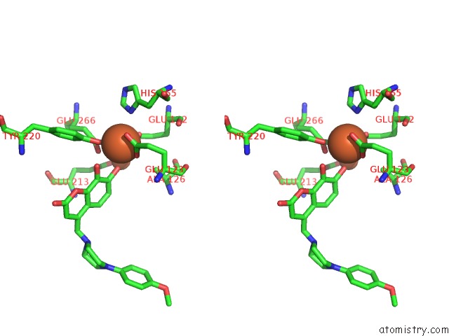
Stereo pair view
Mono view
Stereo pair view
A full contact list of Iron with other atoms in the Fe binding
site number 1 of Crystal Structure of Alternative Oxidase From Trypanosoma Brucei Brucei Complexed with Cumarin Derivative-17 within 5.0Å range:
|
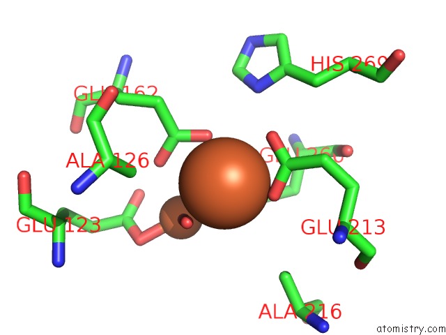
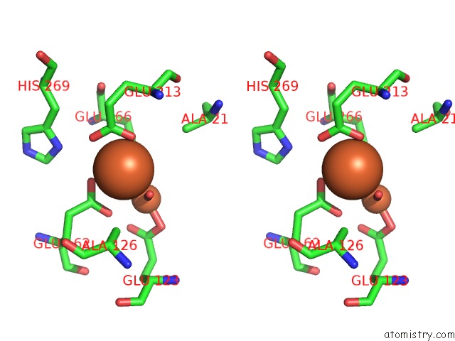
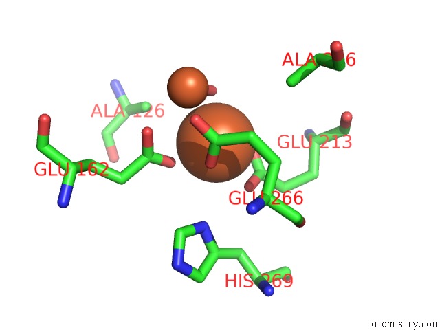
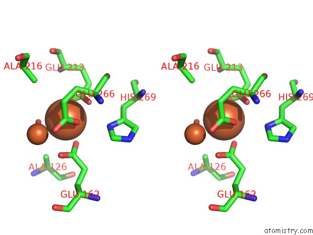
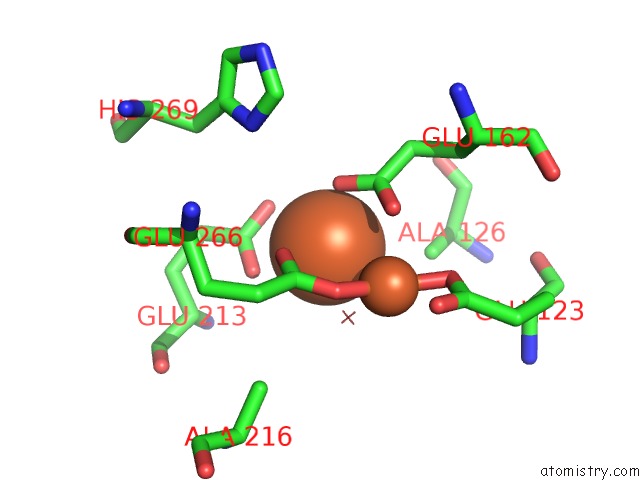
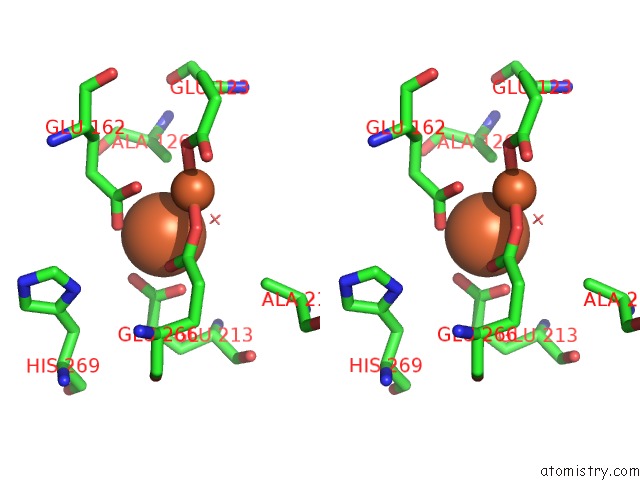
Iron binding site 2 out of 8 in 5gn7
Go back to
Iron binding site 2 out
of 8 in the Crystal Structure of Alternative Oxidase From Trypanosoma Brucei Brucei Complexed with Cumarin Derivative-17
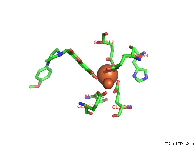
Mono view
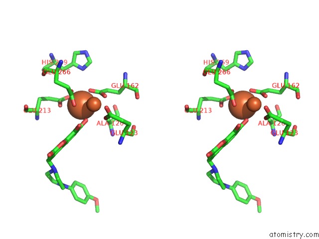
Stereo pair view
Mono view
Stereo pair view
A full contact list of Iron with other atoms in the Fe binding
site number 2 of Crystal Structure of Alternative Oxidase From Trypanosoma Brucei Brucei Complexed with Cumarin Derivative-17 within 5.0Å range:
|
Iron binding site 3 out of 8 in 5gn7
Go back to
Iron binding site 3 out
of 8 in the Crystal Structure of Alternative Oxidase From Trypanosoma Brucei Brucei Complexed with Cumarin Derivative-17
Mono view
Stereo pair view
Mono view
Stereo pair view
A full contact list of Iron with other atoms in the Fe binding
site number 3 of Crystal Structure of Alternative Oxidase From Trypanosoma Brucei Brucei Complexed with Cumarin Derivative-17 within 5.0Å range:
|
Iron binding site 4 out of 8 in 5gn7
Go back to
Iron binding site 4 out
of 8 in the Crystal Structure of Alternative Oxidase From Trypanosoma Brucei Brucei Complexed with Cumarin Derivative-17
Mono view
Stereo pair view
Mono view
Stereo pair view
A full contact list of Iron with other atoms in the Fe binding
site number 4 of Crystal Structure of Alternative Oxidase From Trypanosoma Brucei Brucei Complexed with Cumarin Derivative-17 within 5.0Å range:
|
Iron binding site 5 out of 8 in 5gn7
Go back to
Iron binding site 5 out
of 8 in the Crystal Structure of Alternative Oxidase From Trypanosoma Brucei Brucei Complexed with Cumarin Derivative-17
Mono view
Stereo pair view
Mono view
Stereo pair view
A full contact list of Iron with other atoms in the Fe binding
site number 5 of Crystal Structure of Alternative Oxidase From Trypanosoma Brucei Brucei Complexed with Cumarin Derivative-17 within 5.0Å range:
|
Iron binding site 6 out of 8 in 5gn7
Go back to
Iron binding site 6 out
of 8 in the Crystal Structure of Alternative Oxidase From Trypanosoma Brucei Brucei Complexed with Cumarin Derivative-17
Mono view
Stereo pair view
Mono view
Stereo pair view
A full contact list of Iron with other atoms in the Fe binding
site number 6 of Crystal Structure of Alternative Oxidase From Trypanosoma Brucei Brucei Complexed with Cumarin Derivative-17 within 5.0Å range:
|
Iron binding site 7 out of 8 in 5gn7
Go back to
Iron binding site 7 out
of 8 in the Crystal Structure of Alternative Oxidase From Trypanosoma Brucei Brucei Complexed with Cumarin Derivative-17
Mono view
Stereo pair view
Mono view
Stereo pair view
A full contact list of Iron with other atoms in the Fe binding
site number 7 of Crystal Structure of Alternative Oxidase From Trypanosoma Brucei Brucei Complexed with Cumarin Derivative-17 within 5.0Å range:
|
Iron binding site 8 out of 8 in 5gn7
Go back to
Iron binding site 8 out
of 8 in the Crystal Structure of Alternative Oxidase From Trypanosoma Brucei Brucei Complexed with Cumarin Derivative-17
Mono view
Stereo pair view
Mono view
Stereo pair view
A full contact list of Iron with other atoms in the Fe binding
site number 8 of Crystal Structure of Alternative Oxidase From Trypanosoma Brucei Brucei Complexed with Cumarin Derivative-17 within 5.0Å range:
|
Reference:
E.O.Balogun,
D.K.Inaoka,
T.Shiba,
T.Tsuge,
B.May,
T.Sato,
Y.Kido,
N.Takeshi,
T.Aoki,
T.Honma,
A.Tanaka,
M.Inoue,
S.Matsuoka,
P.A.M.Michels,
Y.Watanabe,
A.L.Moore,
S.Harada,
K.Kita.
Discovery of Trypanocidal Coumarins with Dual-Inhibition For African Trypanosomes Glycerol Kinase and Alternative Oxidase To Be Published.
Page generated: Tue Aug 6 01:33:55 2024
Last articles
F in 7N93F in 7NBK
F in 7NAJ
F in 7N91
F in 7N8R
F in 7N77
F in 7N7W
F in 7N73
F in 7N7L
F in 7N72