Iron »
PDB 5g6f-5h8y »
5gt2 »
Iron in PDB 5gt2: Crystal Structure and Biochemical Features of Dye-Decolorizing Peroxidase Yfex From Escherichia Coli O157
Protein crystallography data
The structure of Crystal Structure and Biochemical Features of Dye-Decolorizing Peroxidase Yfex From Escherichia Coli O157, PDB code: 5gt2
was solved by
Y.L.Ma,
Z.G.Yuan,
S.Liu,
J.X.Wang,
L.C.Gu,
X.H.Liu,
with X-Ray Crystallography technique. A brief refinement statistics is given in the table below:
| Resolution Low / High (Å) | 36.69 / 2.09 |
| Space group | C 1 2 1 |
| Cell size a, b, c (Å), α, β, γ (°) | 121.574, 101.597, 114.300, 90.00, 111.51, 90.00 |
| R / Rfree (%) | 22 / 24.8 |
Iron Binding Sites:
The binding sites of Iron atom in the Crystal Structure and Biochemical Features of Dye-Decolorizing Peroxidase Yfex From Escherichia Coli O157
(pdb code 5gt2). This binding sites where shown within
5.0 Angstroms radius around Iron atom.
In total 4 binding sites of Iron where determined in the Crystal Structure and Biochemical Features of Dye-Decolorizing Peroxidase Yfex From Escherichia Coli O157, PDB code: 5gt2:
Jump to Iron binding site number: 1; 2; 3; 4;
In total 4 binding sites of Iron where determined in the Crystal Structure and Biochemical Features of Dye-Decolorizing Peroxidase Yfex From Escherichia Coli O157, PDB code: 5gt2:
Jump to Iron binding site number: 1; 2; 3; 4;
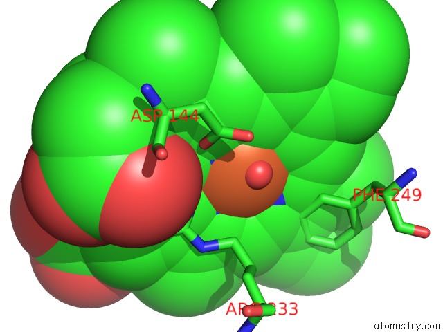
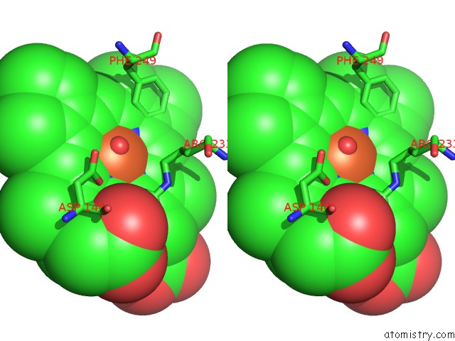
Iron binding site 1 out of 4 in 5gt2
Go back to
Iron binding site 1 out
of 4 in the Crystal Structure and Biochemical Features of Dye-Decolorizing Peroxidase Yfex From Escherichia Coli O157
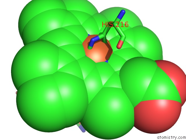
Mono view
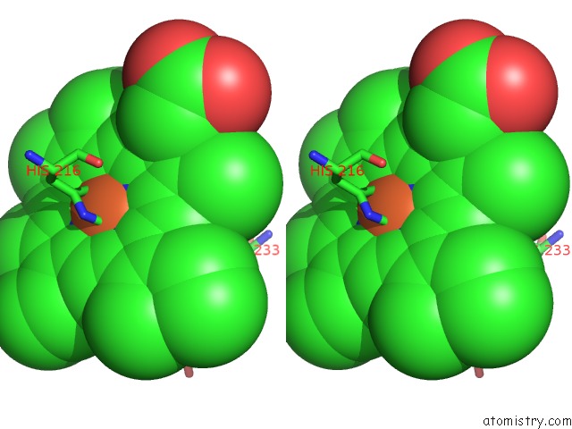
Stereo pair view
Mono view
Stereo pair view
A full contact list of Iron with other atoms in the Fe binding
site number 1 of Crystal Structure and Biochemical Features of Dye-Decolorizing Peroxidase Yfex From Escherichia Coli O157 within 5.0Å range:
|
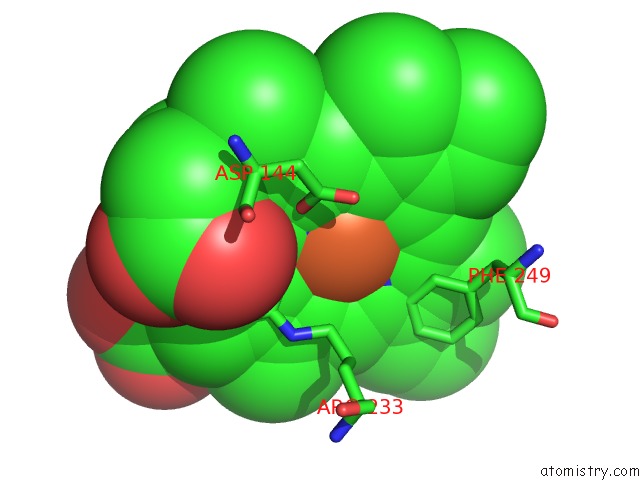
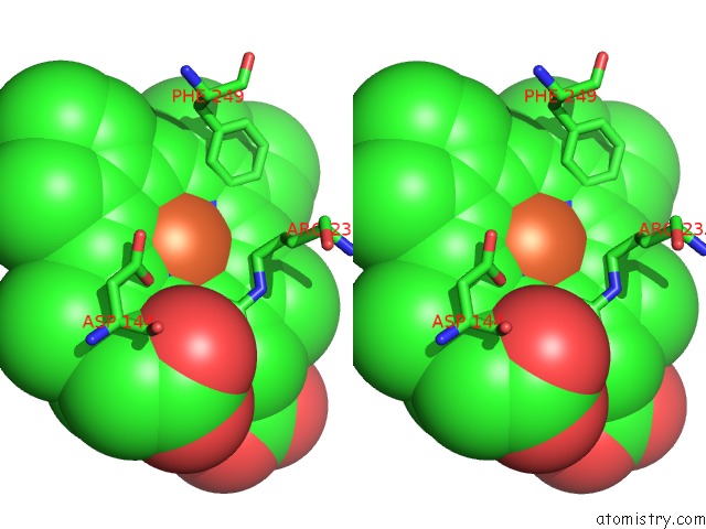
Iron binding site 2 out of 4 in 5gt2
Go back to
Iron binding site 2 out
of 4 in the Crystal Structure and Biochemical Features of Dye-Decolorizing Peroxidase Yfex From Escherichia Coli O157
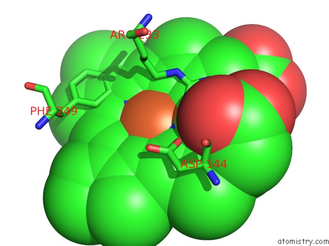
Mono view
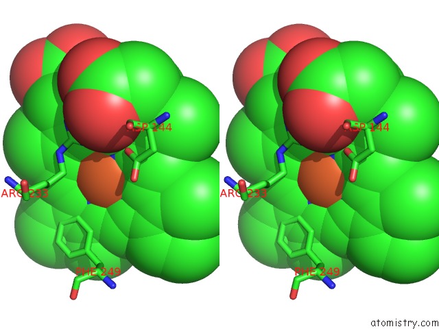
Stereo pair view
Mono view
Stereo pair view
A full contact list of Iron with other atoms in the Fe binding
site number 2 of Crystal Structure and Biochemical Features of Dye-Decolorizing Peroxidase Yfex From Escherichia Coli O157 within 5.0Å range:
|
Iron binding site 3 out of 4 in 5gt2
Go back to
Iron binding site 3 out
of 4 in the Crystal Structure and Biochemical Features of Dye-Decolorizing Peroxidase Yfex From Escherichia Coli O157
Mono view
Stereo pair view
Mono view
Stereo pair view
A full contact list of Iron with other atoms in the Fe binding
site number 3 of Crystal Structure and Biochemical Features of Dye-Decolorizing Peroxidase Yfex From Escherichia Coli O157 within 5.0Å range:
|
Iron binding site 4 out of 4 in 5gt2
Go back to
Iron binding site 4 out
of 4 in the Crystal Structure and Biochemical Features of Dye-Decolorizing Peroxidase Yfex From Escherichia Coli O157
Mono view
Stereo pair view
Mono view
Stereo pair view
A full contact list of Iron with other atoms in the Fe binding
site number 4 of Crystal Structure and Biochemical Features of Dye-Decolorizing Peroxidase Yfex From Escherichia Coli O157 within 5.0Å range:
|
Reference:
X.Liu,
Z.Yuan,
J.Wang,
Y.Cui,
S.Liu,
Y.Ma,
L.Gu,
S.Xu.
Crystal Structure and Biochemical Features of Dye-Decolorizing Peroxidase Yfex From Escherichia Coli O157 Asp(143) and Arg(232) Play Divergent Roles Toward Different Substrates Biochem. Biophys. Res. V. 484 40 2017COMMUN..
ISSN: ESSN 1090-2104
PubMed: 28109884
DOI: 10.1016/J.BBRC.2017.01.081
Page generated: Tue Aug 6 01:34:44 2024
ISSN: ESSN 1090-2104
PubMed: 28109884
DOI: 10.1016/J.BBRC.2017.01.081
Last articles
Zn in 9MJ5Zn in 9HNW
Zn in 9G0L
Zn in 9FNE
Zn in 9DZN
Zn in 9E0I
Zn in 9D32
Zn in 9DAK
Zn in 8ZXC
Zn in 8ZUF