Iron »
PDB 5g6f-5h8y »
5gxg »
Iron in PDB 5gxg: High-Resolution Crystal Structure of the Electron Transfer Complex of Cytochrome P450CAM with Putidaredoxin
Enzymatic activity of High-Resolution Crystal Structure of the Electron Transfer Complex of Cytochrome P450CAM with Putidaredoxin
All present enzymatic activity of High-Resolution Crystal Structure of the Electron Transfer Complex of Cytochrome P450CAM with Putidaredoxin:
1.14.15.1;
1.14.15.1;
Protein crystallography data
The structure of High-Resolution Crystal Structure of the Electron Transfer Complex of Cytochrome P450CAM with Putidaredoxin, PDB code: 5gxg
was solved by
Y.Kikui,
Y.Hiruma,
M.Ubbink,
M.Nojiri,
with X-Ray Crystallography technique. A brief refinement statistics is given in the table below:
| Resolution Low / High (Å) | 59.66 / 1.70 |
| Space group | C 1 2 1 |
| Cell size a, b, c (Å), α, β, γ (°) | 101.490, 77.956, 59.968, 90.00, 95.51, 90.00 |
| R / Rfree (%) | 15.6 / 19.2 |
Iron Binding Sites:
The binding sites of Iron atom in the High-Resolution Crystal Structure of the Electron Transfer Complex of Cytochrome P450CAM with Putidaredoxin
(pdb code 5gxg). This binding sites where shown within
5.0 Angstroms radius around Iron atom.
In total 3 binding sites of Iron where determined in the High-Resolution Crystal Structure of the Electron Transfer Complex of Cytochrome P450CAM with Putidaredoxin, PDB code: 5gxg:
Jump to Iron binding site number: 1; 2; 3;
In total 3 binding sites of Iron where determined in the High-Resolution Crystal Structure of the Electron Transfer Complex of Cytochrome P450CAM with Putidaredoxin, PDB code: 5gxg:
Jump to Iron binding site number: 1; 2; 3;
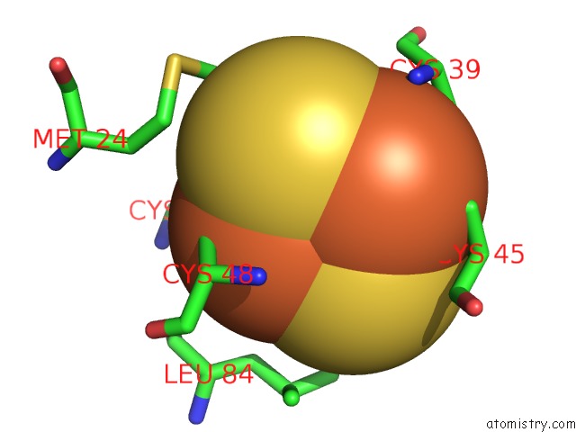
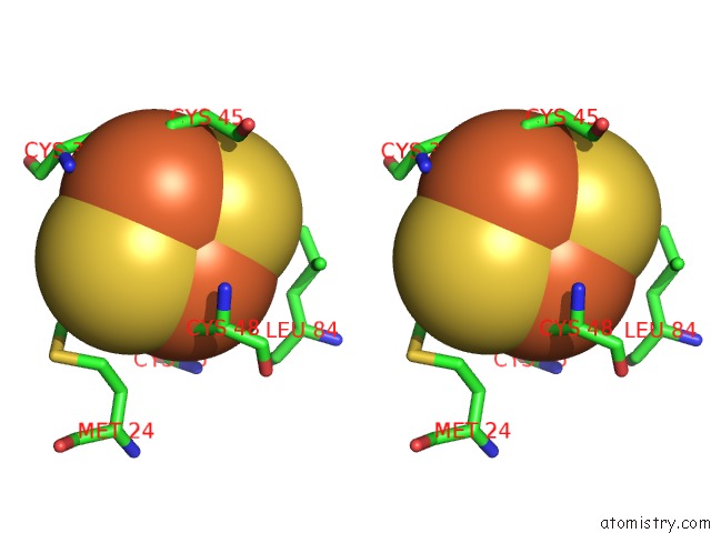
Iron binding site 1 out of 3 in 5gxg
Go back to
Iron binding site 1 out
of 3 in the High-Resolution Crystal Structure of the Electron Transfer Complex of Cytochrome P450CAM with Putidaredoxin
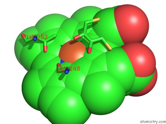
Mono view
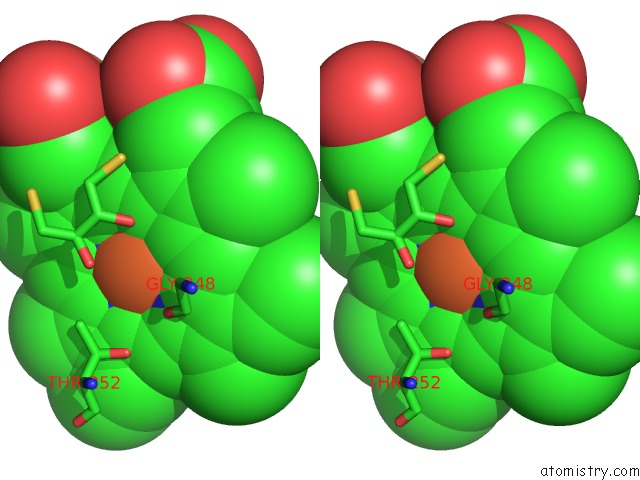
Stereo pair view
Mono view
Stereo pair view
A full contact list of Iron with other atoms in the Fe binding
site number 1 of High-Resolution Crystal Structure of the Electron Transfer Complex of Cytochrome P450CAM with Putidaredoxin within 5.0Å range:
|
Iron binding site 2 out of 3 in 5gxg
Go back to
Iron binding site 2 out
of 3 in the High-Resolution Crystal Structure of the Electron Transfer Complex of Cytochrome P450CAM with Putidaredoxin
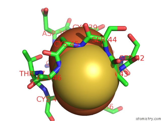
Mono view
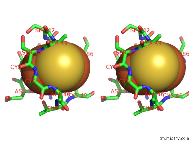
Stereo pair view
Mono view
Stereo pair view
A full contact list of Iron with other atoms in the Fe binding
site number 2 of High-Resolution Crystal Structure of the Electron Transfer Complex of Cytochrome P450CAM with Putidaredoxin within 5.0Å range:
|
Iron binding site 3 out of 3 in 5gxg
Go back to
Iron binding site 3 out
of 3 in the High-Resolution Crystal Structure of the Electron Transfer Complex of Cytochrome P450CAM with Putidaredoxin
Mono view
Stereo pair view
Mono view
Stereo pair view
A full contact list of Iron with other atoms in the Fe binding
site number 3 of High-Resolution Crystal Structure of the Electron Transfer Complex of Cytochrome P450CAM with Putidaredoxin within 5.0Å range:
|
Reference:
W.Andraojc,
Y.Hiruma,
W.M.Liu,
E.Ravera,
M.Nojiri,
G.Parigi,
C.Luchinat,
M.Ubbink.
Identification of Productive and Futile Encounters in An Electron Transfer Protein Complex Proc. Natl. Acad. Sci. V. 114 E1840 2017U.S.A..
ISSN: ESSN 1091-6490
PubMed: 28223532
DOI: 10.1073/PNAS.1616813114
Page generated: Tue Aug 6 01:41:00 2024
ISSN: ESSN 1091-6490
PubMed: 28223532
DOI: 10.1073/PNAS.1616813114
Last articles
Zn in 9MJ5Zn in 9HNW
Zn in 9G0L
Zn in 9FNE
Zn in 9DZN
Zn in 9E0I
Zn in 9D32
Zn in 9DAK
Zn in 8ZXC
Zn in 8ZUF