Iron »
PDB 5g6f-5h8y »
5h1z »
Iron in PDB 5h1z: CYP153D17 From Sphingomonas Sp. Pamc 26605
Protein crystallography data
The structure of CYP153D17 From Sphingomonas Sp. Pamc 26605, PDB code: 5h1z
was solved by
C.W.Lee,
J.H.Lee,
with X-Ray Crystallography technique. A brief refinement statistics is given in the table below:
| Resolution Low / High (Å) | 50.01 / 3.10 |
| Space group | P 31 2 1 |
| Cell size a, b, c (Å), α, β, γ (°) | 110.837, 110.837, 113.180, 90.00, 90.00, 120.00 |
| R / Rfree (%) | 26.2 / 34 |
Iron Binding Sites:
The binding sites of Iron atom in the CYP153D17 From Sphingomonas Sp. Pamc 26605
(pdb code 5h1z). This binding sites where shown within
5.0 Angstroms radius around Iron atom.
In total only one binding site of Iron was determined in the CYP153D17 From Sphingomonas Sp. Pamc 26605, PDB code: 5h1z:
In total only one binding site of Iron was determined in the CYP153D17 From Sphingomonas Sp. Pamc 26605, PDB code: 5h1z:
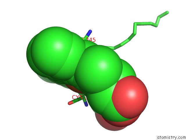
Iron binding site 1 out of 1 in 5h1z
Go back to
Iron binding site 1 out
of 1 in the CYP153D17 From Sphingomonas Sp. Pamc 26605
Mono view

Stereo pair view
Mono view
Stereo pair view
A full contact list of Iron with other atoms in the Fe binding
site number 1 of CYP153D17 From Sphingomonas Sp. Pamc 26605 within 5.0Å range:
|
Reference:
C.W.Lee,
S.C.Yu,
J.H.Lee,
S.H.Park,
H.Park,
T.J.Oh,
J.H.Lee.
Crystal Structure of A Putative Cytochrome P450 Alkane Hydroxylase (CYP153D17) From Sphingomonas Sp. Pamc 26605 and Its Conformational Substrate Binding Int J Mol Sci V. 17 2016.
ISSN: ESSN 1422-0067
PubMed: 27941697
DOI: 10.3390/IJMS17122067
Page generated: Tue Aug 6 01:41:02 2024
ISSN: ESSN 1422-0067
PubMed: 27941697
DOI: 10.3390/IJMS17122067
Last articles
Fe in 2YXOFe in 2YRS
Fe in 2YXC
Fe in 2YNM
Fe in 2YVJ
Fe in 2YP1
Fe in 2YU2
Fe in 2YU1
Fe in 2YQB
Fe in 2YOO