Iron »
PDB 5g6f-5h8y »
5h8c »
Iron in PDB 5h8c: Truncated Xpd
Protein crystallography data
The structure of Truncated Xpd, PDB code: 5h8c
was solved by
J.H.Naismith,
D.Constantinescu,
with X-Ray Crystallography technique. A brief refinement statistics is given in the table below:
| Resolution Low / High (Å) | 29.00 / 2.29 |
| Space group | P 21 21 21 |
| Cell size a, b, c (Å), α, β, γ (°) | 64.800, 77.800, 99.540, 90.00, 90.00, 90.00 |
| R / Rfree (%) | 20.3 / 23.9 |
Iron Binding Sites:
The binding sites of Iron atom in the Truncated Xpd
(pdb code 5h8c). This binding sites where shown within
5.0 Angstroms radius around Iron atom.
In total 4 binding sites of Iron where determined in the Truncated Xpd, PDB code: 5h8c:
Jump to Iron binding site number: 1; 2; 3; 4;
In total 4 binding sites of Iron where determined in the Truncated Xpd, PDB code: 5h8c:
Jump to Iron binding site number: 1; 2; 3; 4;
Iron binding site 1 out of 4 in 5h8c
Go back to
Iron binding site 1 out
of 4 in the Truncated Xpd
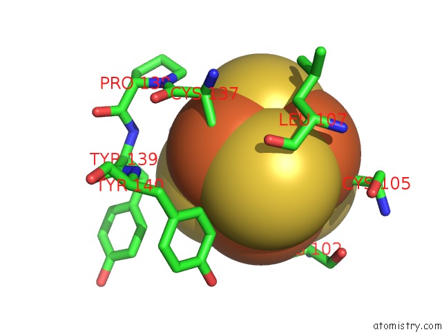
Mono view
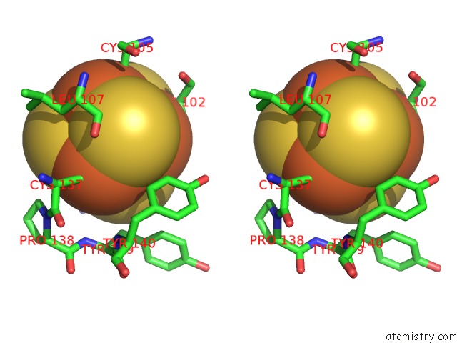
Stereo pair view
Mono view
Stereo pair view
A full contact list of Iron with other atoms in the Fe binding
site number 1 of Truncated Xpd within 5.0Å range:
|
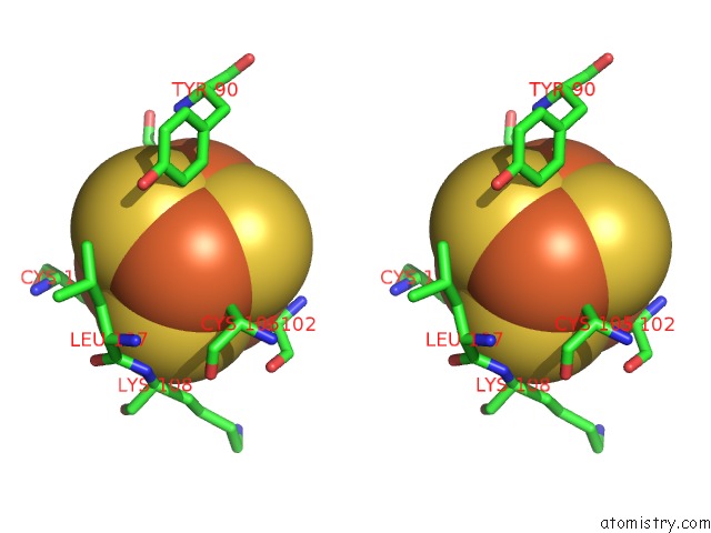
Iron binding site 2 out of 4 in 5h8c
Go back to
Iron binding site 2 out
of 4 in the Truncated Xpd

Mono view

Stereo pair view
Mono view
Stereo pair view
A full contact list of Iron with other atoms in the Fe binding
site number 2 of Truncated Xpd within 5.0Å range:
|
Iron binding site 3 out of 4 in 5h8c
Go back to
Iron binding site 3 out
of 4 in the Truncated Xpd

Mono view

Stereo pair view
Mono view
Stereo pair view
A full contact list of Iron with other atoms in the Fe binding
site number 3 of Truncated Xpd within 5.0Å range:
|
Iron binding site 4 out of 4 in 5h8c
Go back to
Iron binding site 4 out
of 4 in the Truncated Xpd

Mono view
Stereo pair view
Mono view
Stereo pair view
A full contact list of Iron with other atoms in the Fe binding
site number 4 of Truncated Xpd within 5.0Å range:
|
Reference:
D.Constantinescu-Aruxandei,
B.Petrovic-Stojanovska,
J.C.Penedo,
M.F.White,
J.H.Naismith.
Mechanism of Dna Loading By the Dna Repair Helicase Xpd. Nucleic Acids Res. V. 44 2806 2016.
ISSN: ESSN 1362-4962
PubMed: 26896802
DOI: 10.1093/NAR/GKW102
Page generated: Tue Aug 6 01:43:39 2024
ISSN: ESSN 1362-4962
PubMed: 26896802
DOI: 10.1093/NAR/GKW102
Last articles
Zn in 9MJ5Zn in 9HNW
Zn in 9G0L
Zn in 9FNE
Zn in 9DZN
Zn in 9E0I
Zn in 9D32
Zn in 9DAK
Zn in 8ZXC
Zn in 8ZUF