Iron »
PDB 5ibf-5j84 »
5j6d »
Iron in PDB 5j6d: Discovery of Acyl Guanidine Tryptophan Hydroxylase-1 Inhibitors
Enzymatic activity of Discovery of Acyl Guanidine Tryptophan Hydroxylase-1 Inhibitors
All present enzymatic activity of Discovery of Acyl Guanidine Tryptophan Hydroxylase-1 Inhibitors:
1.14.16.4;
1.14.16.4;
Protein crystallography data
The structure of Discovery of Acyl Guanidine Tryptophan Hydroxylase-1 Inhibitors, PDB code: 5j6d
was solved by
A.J.Stein,
D.R.Goldberg,
S.De Lombaert,
with X-Ray Crystallography technique. A brief refinement statistics is given in the table below:
| Resolution Low / High (Å) | 50.00 / 1.90 |
| Space group | P 21 21 21 |
| Cell size a, b, c (Å), α, β, γ (°) | 58.726, 63.561, 156.468, 90.00, 90.00, 90.00 |
| R / Rfree (%) | 21 / 24.8 |
Iron Binding Sites:
The binding sites of Iron atom in the Discovery of Acyl Guanidine Tryptophan Hydroxylase-1 Inhibitors
(pdb code 5j6d). This binding sites where shown within
5.0 Angstroms radius around Iron atom.
In total 2 binding sites of Iron where determined in the Discovery of Acyl Guanidine Tryptophan Hydroxylase-1 Inhibitors, PDB code: 5j6d:
Jump to Iron binding site number: 1; 2;
In total 2 binding sites of Iron where determined in the Discovery of Acyl Guanidine Tryptophan Hydroxylase-1 Inhibitors, PDB code: 5j6d:
Jump to Iron binding site number: 1; 2;
Iron binding site 1 out of 2 in 5j6d
Go back to
Iron binding site 1 out
of 2 in the Discovery of Acyl Guanidine Tryptophan Hydroxylase-1 Inhibitors
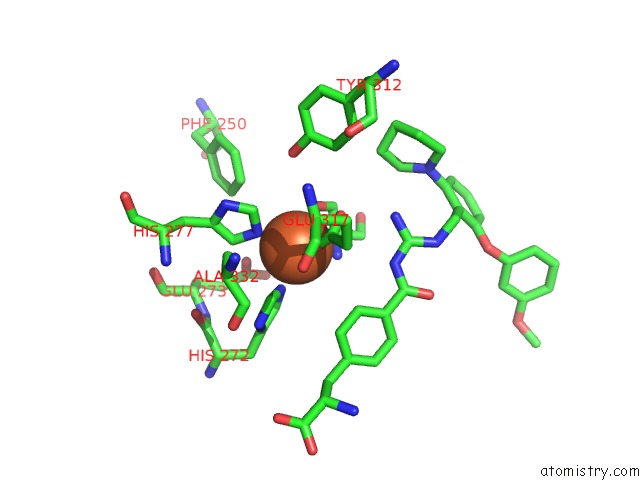
Mono view
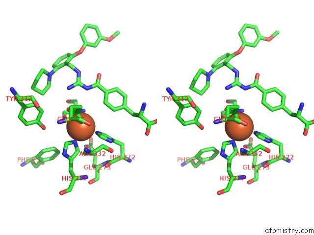
Stereo pair view
Mono view
Stereo pair view
A full contact list of Iron with other atoms in the Fe binding
site number 1 of Discovery of Acyl Guanidine Tryptophan Hydroxylase-1 Inhibitors within 5.0Å range:
|
Iron binding site 2 out of 2 in 5j6d
Go back to
Iron binding site 2 out
of 2 in the Discovery of Acyl Guanidine Tryptophan Hydroxylase-1 Inhibitors
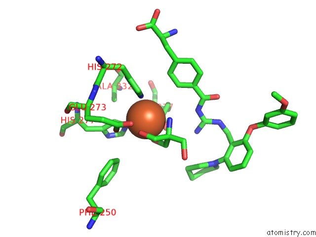
Mono view
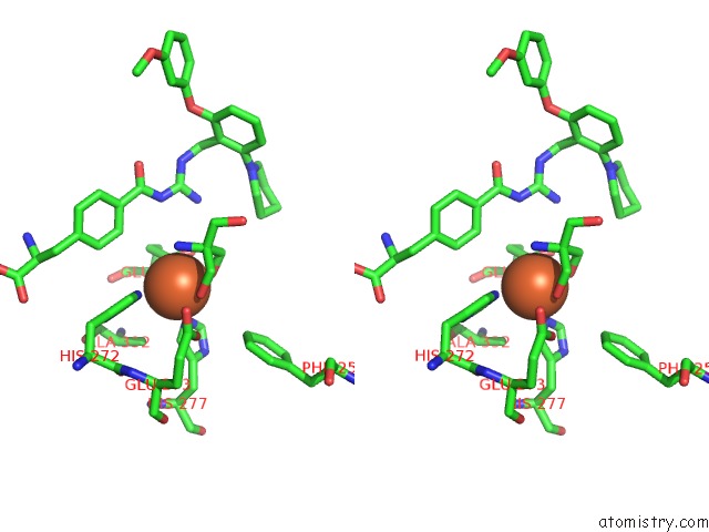
Stereo pair view
Mono view
Stereo pair view
A full contact list of Iron with other atoms in the Fe binding
site number 2 of Discovery of Acyl Guanidine Tryptophan Hydroxylase-1 Inhibitors within 5.0Å range:
|
Reference:
D.R.Goldberg,
S.De Lombaert,
R.Aiello,
P.Bourassa,
N.Barucci,
Q.Zhang,
V.Paralkar,
A.J.Stein,
J.Valentine,
W.Zavadoski.
Discovery of Acyl Guanidine Tryptophan Hydroxylase-1 Inhibitors. Bioorg.Med.Chem.Lett. V. 26 2855 2016.
ISSN: ESSN 1464-3405
PubMed: 27146606
DOI: 10.1016/J.BMCL.2016.04.057
Page generated: Tue Aug 6 02:22:55 2024
ISSN: ESSN 1464-3405
PubMed: 27146606
DOI: 10.1016/J.BMCL.2016.04.057
Last articles
Zn in 9JYWZn in 9IR4
Zn in 9IR3
Zn in 9GMX
Zn in 9GMW
Zn in 9JEJ
Zn in 9ERF
Zn in 9ERE
Zn in 9EGV
Zn in 9EGW