Iron »
PDB 5o17-5ok4 »
5ojb »
Iron in PDB 5ojb: Structure of Mbq Nmh
Protein crystallography data
The structure of Structure of Mbq Nmh, PDB code: 5ojb
was solved by
T.Hayashi,
M.Pott,
T.Mori,
P.Mittl,
A.Green,
D.Hivert,
with X-Ray Crystallography technique. A brief refinement statistics is given in the table below:
| Resolution Low / High (Å) | 45.57 / 1.54 |
| Space group | P 65 |
| Cell size a, b, c (Å), α, β, γ (°) | 91.134, 91.134, 39.687, 90.00, 90.00, 120.00 |
| R / Rfree (%) | 20.1 / 23.3 |
Iron Binding Sites:
The binding sites of Iron atom in the Structure of Mbq Nmh
(pdb code 5ojb). This binding sites where shown within
5.0 Angstroms radius around Iron atom.
In total only one binding site of Iron was determined in the Structure of Mbq Nmh, PDB code: 5ojb:
In total only one binding site of Iron was determined in the Structure of Mbq Nmh, PDB code: 5ojb:
Iron binding site 1 out of 1 in 5ojb
Go back to
Iron binding site 1 out
of 1 in the Structure of Mbq Nmh
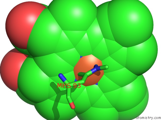
Mono view
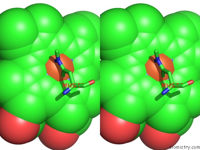
Stereo pair view
Mono view
Stereo pair view
A full contact list of Iron with other atoms in the Fe binding
site number 1 of Structure of Mbq Nmh within 5.0Å range:
|
Reference:
M.Pott,
T.Hayashi,
T.Mori,
P.R.E.Mittl,
A.P.Green,
D.Hilvert.
A Noncanonical Proximal Heme Ligand Affords An Efficient Peroxidase in A Globin Fold. J. Am. Chem. Soc. V. 140 1535 2018.
ISSN: ESSN 1520-5126
PubMed: 29309143
DOI: 10.1021/JACS.7B12621
Page generated: Wed Aug 6 01:05:15 2025
ISSN: ESSN 1520-5126
PubMed: 29309143
DOI: 10.1021/JACS.7B12621
Last articles
Fe in 6ZI2Fe in 6ZI3
Fe in 6ZH5
Fe in 6ZHZ
Fe in 6ZHW
Fe in 6ZCE
Fe in 6ZFU
Fe in 6ZFS
Fe in 6ZFT
Fe in 6ZAQ