Iron »
PDB 5xdh-5xy4 »
5xe1 »
Iron in PDB 5xe1: Crystal Structure of the Indoleamine 2,3-Dioxygenagse 1 (IDO1) Complexed with INCB14943
Enzymatic activity of Crystal Structure of the Indoleamine 2,3-Dioxygenagse 1 (IDO1) Complexed with INCB14943
All present enzymatic activity of Crystal Structure of the Indoleamine 2,3-Dioxygenagse 1 (IDO1) Complexed with INCB14943:
1.13.11.52;
1.13.11.52;
Protein crystallography data
The structure of Crystal Structure of the Indoleamine 2,3-Dioxygenagse 1 (IDO1) Complexed with INCB14943, PDB code: 5xe1
was solved by
J.Xu,
U.Wu,
J.Liu,
with X-Ray Crystallography technique. A brief refinement statistics is given in the table below:
| Resolution Low / High (Å) | 25.00 / 3.20 |
| Space group | P 21 21 21 |
| Cell size a, b, c (Å), α, β, γ (°) | 87.050, 97.380, 128.470, 90.00, 90.00, 90.00 |
| R / Rfree (%) | 22.8 / 27.4 |
Other elements in 5xe1:
The structure of Crystal Structure of the Indoleamine 2,3-Dioxygenagse 1 (IDO1) Complexed with INCB14943 also contains other interesting chemical elements:
| Fluorine | (F) | 2 atoms |
| Chlorine | (Cl) | 2 atoms |
Iron Binding Sites:
The binding sites of Iron atom in the Crystal Structure of the Indoleamine 2,3-Dioxygenagse 1 (IDO1) Complexed with INCB14943
(pdb code 5xe1). This binding sites where shown within
5.0 Angstroms radius around Iron atom.
In total 2 binding sites of Iron where determined in the Crystal Structure of the Indoleamine 2,3-Dioxygenagse 1 (IDO1) Complexed with INCB14943, PDB code: 5xe1:
Jump to Iron binding site number: 1; 2;
In total 2 binding sites of Iron where determined in the Crystal Structure of the Indoleamine 2,3-Dioxygenagse 1 (IDO1) Complexed with INCB14943, PDB code: 5xe1:
Jump to Iron binding site number: 1; 2;
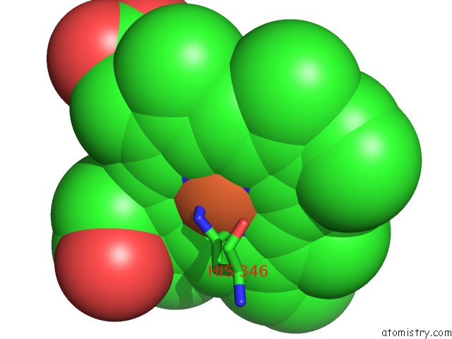
Iron binding site 1 out of 2 in 5xe1
Go back to
Iron binding site 1 out
of 2 in the Crystal Structure of the Indoleamine 2,3-Dioxygenagse 1 (IDO1) Complexed with INCB14943

Mono view

Stereo pair view
Mono view
Stereo pair view
A full contact list of Iron with other atoms in the Fe binding
site number 1 of Crystal Structure of the Indoleamine 2,3-Dioxygenagse 1 (IDO1) Complexed with INCB14943 within 5.0Å range:
|
Iron binding site 2 out of 2 in 5xe1
Go back to
Iron binding site 2 out
of 2 in the Crystal Structure of the Indoleamine 2,3-Dioxygenagse 1 (IDO1) Complexed with INCB14943
Mono view

Stereo pair view
Mono view
Stereo pair view
A full contact list of Iron with other atoms in the Fe binding
site number 2 of Crystal Structure of the Indoleamine 2,3-Dioxygenagse 1 (IDO1) Complexed with INCB14943 within 5.0Å range:
|
Reference:
Y.Wu,
T.Xu,
J.Liu,
K.Ding,
J.Xu.
Structural Insights Into the Binding Mechanism of IDO1 with Hydroxylamidine Based Inhibitor INCB14943 Biochem. Biophys. Res. V. 487 339 2017COMMUN..
ISSN: ESSN 1090-2104
PubMed: 28412361
DOI: 10.1016/J.BBRC.2017.04.061
Page generated: Tue Aug 6 11:39:25 2024
ISSN: ESSN 1090-2104
PubMed: 28412361
DOI: 10.1016/J.BBRC.2017.04.061
Last articles
Zn in 9MJ5Zn in 9HNW
Zn in 9G0L
Zn in 9FNE
Zn in 9DZN
Zn in 9E0I
Zn in 9D32
Zn in 9DAK
Zn in 8ZXC
Zn in 8ZUF