Iron »
PDB 6g71-6gl6 »
6gkc »
Iron in PDB 6gkc: 2 Minute FE2+ Soak Structure of Synftn
Enzymatic activity of 2 Minute FE2+ Soak Structure of Synftn
All present enzymatic activity of 2 Minute FE2+ Soak Structure of Synftn:
1.16.3.2;
1.16.3.2;
Protein crystallography data
The structure of 2 Minute FE2+ Soak Structure of Synftn, PDB code: 6gkc
was solved by
A.M.Hemmings,
J.M.Bradley,
with X-Ray Crystallography technique. A brief refinement statistics is given in the table below:
| Resolution Low / High (Å) | 53.41 / 1.97 |
| Space group | F 4 3 2 |
| Cell size a, b, c (Å), α, β, γ (°) | 177.157, 177.157, 177.157, 90.00, 90.00, 90.00 |
| R / Rfree (%) | 17.8 / 20.1 |
Iron Binding Sites:
The binding sites of Iron atom in the 2 Minute FE2+ Soak Structure of Synftn
(pdb code 6gkc). This binding sites where shown within
5.0 Angstroms radius around Iron atom.
In total 2 binding sites of Iron where determined in the 2 Minute FE2+ Soak Structure of Synftn, PDB code: 6gkc:
Jump to Iron binding site number: 1; 2;
In total 2 binding sites of Iron where determined in the 2 Minute FE2+ Soak Structure of Synftn, PDB code: 6gkc:
Jump to Iron binding site number: 1; 2;
Iron binding site 1 out of 2 in 6gkc
Go back to
Iron binding site 1 out
of 2 in the 2 Minute FE2+ Soak Structure of Synftn
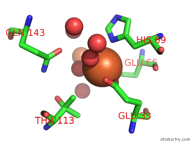
Mono view
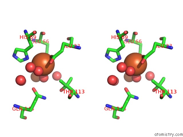
Stereo pair view
Mono view
Stereo pair view
A full contact list of Iron with other atoms in the Fe binding
site number 1 of 2 Minute FE2+ Soak Structure of Synftn within 5.0Å range:
|
Iron binding site 2 out of 2 in 6gkc
Go back to
Iron binding site 2 out
of 2 in the 2 Minute FE2+ Soak Structure of Synftn
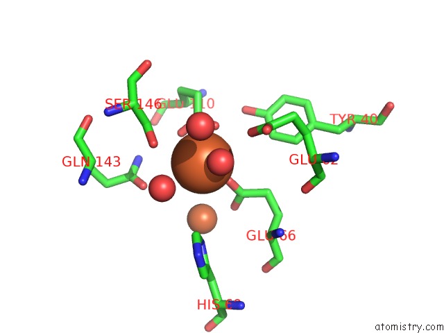
Mono view
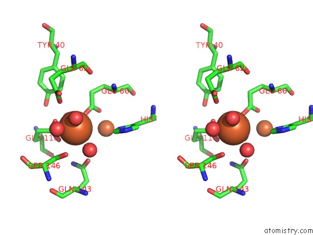
Stereo pair view
Mono view
Stereo pair view
A full contact list of Iron with other atoms in the Fe binding
site number 2 of 2 Minute FE2+ Soak Structure of Synftn within 5.0Å range:
|
Reference:
J.M.Bradley,
D.A.Svistunenko,
J.Pullin,
N.Hill,
R.K.Stuart,
B.Palenik,
M.T.Wilson,
A.M.Hemmings,
G.R.Moore,
N.E.Le Brun.
Reaction of O2WITH A Diiron Protein Generates A Mixed-Valent FE2+/FE3+Center and Peroxide. Proc. Natl. Acad. Sci. V. 116 2058 2019U.S.A..
ISSN: ESSN 1091-6490
PubMed: 30659147
DOI: 10.1073/PNAS.1809913116
Page generated: Wed Aug 6 06:55:14 2025
ISSN: ESSN 1091-6490
PubMed: 30659147
DOI: 10.1073/PNAS.1809913116
Last articles
Fe in 6L86Fe in 6L6S
Fe in 6L6J
Fe in 6L6X
Fe in 6L6W
Fe in 6L5Y
Fe in 6L5X
Fe in 6L69
Fe in 6L55
Fe in 6L5W