Iron »
PDB 6gly-6haw »
6h1h »
Iron in PDB 6h1h: Crystal Structure of Human Pirin in Complex with Compound 7 (PLX4720)
Enzymatic activity of Crystal Structure of Human Pirin in Complex with Compound 7 (PLX4720)
All present enzymatic activity of Crystal Structure of Human Pirin in Complex with Compound 7 (PLX4720):
1.13.11.24;
1.13.11.24;
Protein crystallography data
The structure of Crystal Structure of Human Pirin in Complex with Compound 7 (PLX4720), PDB code: 6h1h
was solved by
S.Ali,
Y.V.Le Bihan,
R.L.M.Van Montfort,
with X-Ray Crystallography technique. A brief refinement statistics is given in the table below:
| Resolution Low / High (Å) | 34.00 / 1.54 |
| Space group | P 21 21 21 |
| Cell size a, b, c (Å), α, β, γ (°) | 42.300, 67.408, 107.681, 90.00, 90.00, 90.00 |
| R / Rfree (%) | 15.9 / 18.3 |
Other elements in 6h1h:
The structure of Crystal Structure of Human Pirin in Complex with Compound 7 (PLX4720) also contains other interesting chemical elements:
| Fluorine | (F) | 4 atoms |
| Chlorine | (Cl) | 2 atoms |
Iron Binding Sites:
The binding sites of Iron atom in the Crystal Structure of Human Pirin in Complex with Compound 7 (PLX4720)
(pdb code 6h1h). This binding sites where shown within
5.0 Angstroms radius around Iron atom.
In total only one binding site of Iron was determined in the Crystal Structure of Human Pirin in Complex with Compound 7 (PLX4720), PDB code: 6h1h:
In total only one binding site of Iron was determined in the Crystal Structure of Human Pirin in Complex with Compound 7 (PLX4720), PDB code: 6h1h:
Iron binding site 1 out of 1 in 6h1h
Go back to
Iron binding site 1 out
of 1 in the Crystal Structure of Human Pirin in Complex with Compound 7 (PLX4720)
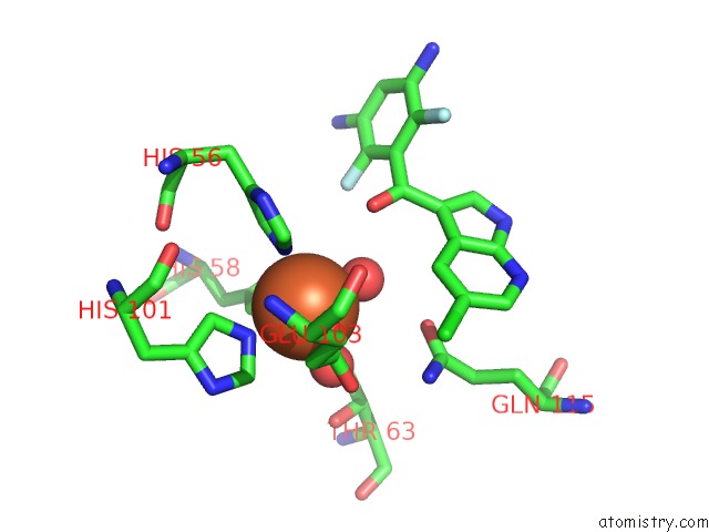
Mono view
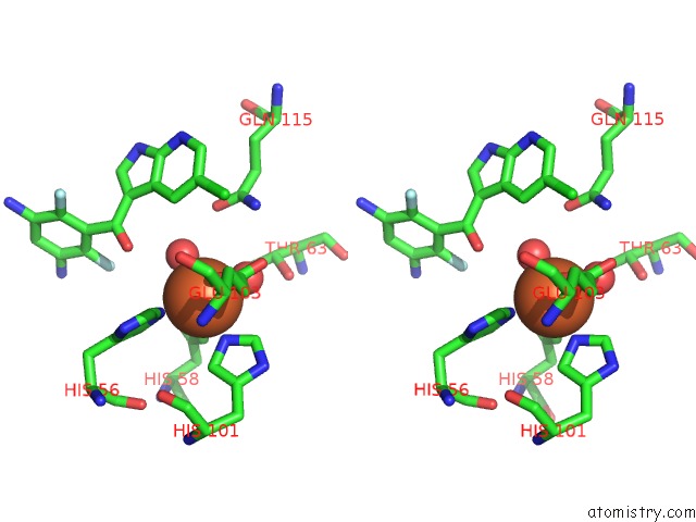
Stereo pair view
Mono view
Stereo pair view
A full contact list of Iron with other atoms in the Fe binding
site number 1 of Crystal Structure of Human Pirin in Complex with Compound 7 (PLX4720) within 5.0Å range:
|
Reference:
J.Meyers,
N.E.A.Chessum,
S.Ali,
N.Y.Mok,
B.Wilding,
A.E.Pasqua,
M.Rowlands,
M.J.Tucker,
L.E.Evans,
C.S.Rye,
L.O'fee,
Y.V.Le Bihan,
R.Burke,
M.Carter,
P.Workman,
J.Blagg,
N.Brown,
R.L.M.Van Montfort,
K.Jones,
M.D.Cheeseman.
Privileged Structures and Polypharmacology Within and Between Protein Families. Acs Med Chem Lett V. 9 1199 2018.
ISSN: ISSN 1948-5875
PubMed: 30613326
DOI: 10.1021/ACSMEDCHEMLETT.8B00364
Page generated: Wed Aug 6 07:27:25 2025
ISSN: ISSN 1948-5875
PubMed: 30613326
DOI: 10.1021/ACSMEDCHEMLETT.8B00364
Last articles
Fe in 6M0AFe in 6LVV
Fe in 6LU1
Fe in 6LY4
Fe in 6LVC
Fe in 6LVB
Fe in 6LS3
Fe in 6LS8
Fe in 6LTM
Fe in 6LTL