Iron »
PDB 6yj4-6zaf »
6z1m »
Iron in PDB 6z1m: Structure of An Ancestral Glycosidase (Family 1) Bound to Heme
Protein crystallography data
The structure of Structure of An Ancestral Glycosidase (Family 1) Bound to Heme, PDB code: 6z1m
was solved by
J.A.Gavira,
V.A.Risso,
J.M.Sanchez-Ruiz,
G.Gamiz-Arco,
L.Gutierrez-Rus,
B.Ibarra-Molero,
Y.Oshino,
D.Petrovic,
A.Romero-Rivera,
B.Seelig,
S.C.L.Kamerlin,
E.A.Gaucher,
with X-Ray Crystallography technique. A brief refinement statistics is given in the table below:
| Resolution Low / High (Å) | 55.32 / 2.45 |
| Space group | P 1 21 1 |
| Cell size a, b, c (Å), α, β, γ (°) | 58.934, 89.496, 141.120, 90.00, 94.21, 90.00 |
| R / Rfree (%) | 17.5 / 22.1 |
Other elements in 6z1m:
The structure of Structure of An Ancestral Glycosidase (Family 1) Bound to Heme also contains other interesting chemical elements:
| Magnesium | (Mg) | 1 atom |
Iron Binding Sites:
The binding sites of Iron atom in the Structure of An Ancestral Glycosidase (Family 1) Bound to Heme
(pdb code 6z1m). This binding sites where shown within
5.0 Angstroms radius around Iron atom.
In total 3 binding sites of Iron where determined in the Structure of An Ancestral Glycosidase (Family 1) Bound to Heme, PDB code: 6z1m:
Jump to Iron binding site number: 1; 2; 3;
In total 3 binding sites of Iron where determined in the Structure of An Ancestral Glycosidase (Family 1) Bound to Heme, PDB code: 6z1m:
Jump to Iron binding site number: 1; 2; 3;
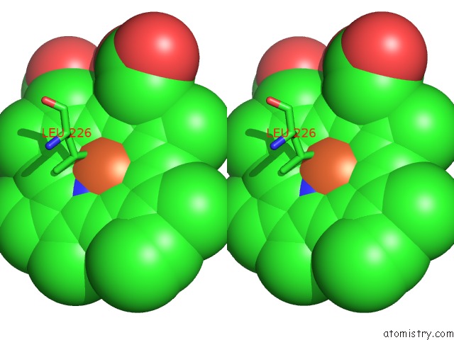
Iron binding site 1 out of 3 in 6z1m
Go back to
Iron binding site 1 out
of 3 in the Structure of An Ancestral Glycosidase (Family 1) Bound to Heme
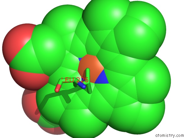
Mono view
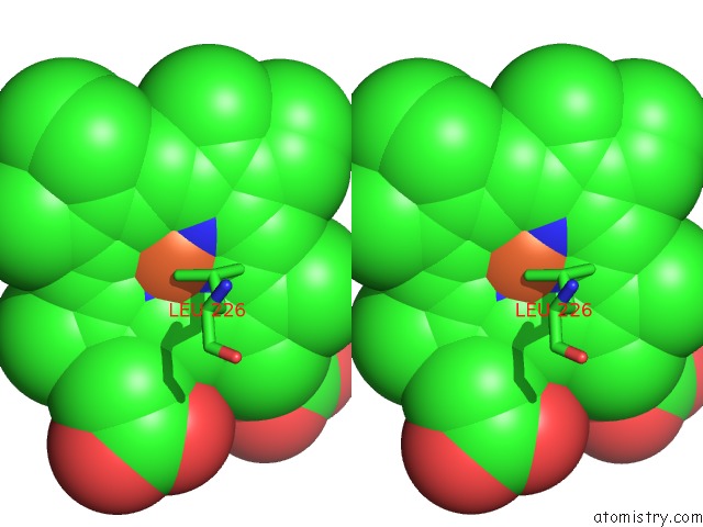
Stereo pair view
Mono view
Stereo pair view
A full contact list of Iron with other atoms in the Fe binding
site number 1 of Structure of An Ancestral Glycosidase (Family 1) Bound to Heme within 5.0Å range:
|
Iron binding site 2 out of 3 in 6z1m
Go back to
Iron binding site 2 out
of 3 in the Structure of An Ancestral Glycosidase (Family 1) Bound to Heme
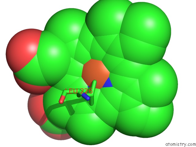
Mono view

Stereo pair view
Mono view
Stereo pair view
A full contact list of Iron with other atoms in the Fe binding
site number 2 of Structure of An Ancestral Glycosidase (Family 1) Bound to Heme within 5.0Å range:
|
Iron binding site 3 out of 3 in 6z1m
Go back to
Iron binding site 3 out
of 3 in the Structure of An Ancestral Glycosidase (Family 1) Bound to Heme
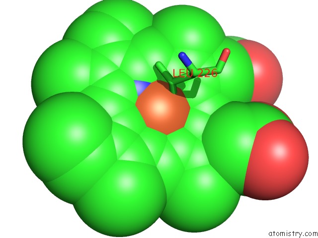
Mono view
Stereo pair view
Mono view
Stereo pair view
A full contact list of Iron with other atoms in the Fe binding
site number 3 of Structure of An Ancestral Glycosidase (Family 1) Bound to Heme within 5.0Å range:
|
Reference:
G.Gamiz-Arco,
L.Gutierrez-Rus,
V.A.Risso,
B.Ibarra-Molero,
Y.Oshino,
D.Petrovic,
A.Romero-Rivera,
B.Seelig,
J.A.Gavira,
S.C.L.Kamerlin,
E.A.Gaucher,
J.M.Sanchez-Ruiz.
Novel Heme-Binding Enables Allosteric Modulation in An Ancient Tim-Barrel Glycosidase Biorxiv 2020.
DOI: 10.1101/2020.05.27.118968
Page generated: Wed Aug 7 16:53:53 2024
DOI: 10.1101/2020.05.27.118968
Last articles
Cl in 3BMBCl in 3BMY
Cl in 3BMW
Cl in 3BLR
Cl in 3BLP
Cl in 3BLK
Cl in 3BLA
Cl in 3BL9
Cl in 3BLJ
Cl in 3BL1