Iron »
PDB 7ai9-7bhb »
7bbt »
Iron in PDB 7bbt: Structure of Cytochrome C in Complex with A P-Benzyl-Sulfonato- Calix[8]Arene-Peg Pseudorotaxane
Protein crystallography data
The structure of Structure of Cytochrome C in Complex with A P-Benzyl-Sulfonato- Calix[8]Arene-Peg Pseudorotaxane, PDB code: 7bbt
was solved by
N.M.Mockler,
K.Ramberg,
F.Guagnini,
C.L.Raston,
P.B.Crowley,
with X-Ray Crystallography technique. A brief refinement statistics is given in the table below:
| Resolution Low / High (Å) | 50.57 / 3.02 |
| Space group | C 1 2 1 |
| Cell size a, b, c (Å), α, β, γ (°) | 120.851, 70.215, 70.175, 90, 102.93, 90 |
| R / Rfree (%) | 23.3 / 26.1 |
Iron Binding Sites:
The binding sites of Iron atom in the Structure of Cytochrome C in Complex with A P-Benzyl-Sulfonato- Calix[8]Arene-Peg Pseudorotaxane
(pdb code 7bbt). This binding sites where shown within
5.0 Angstroms radius around Iron atom.
In total 4 binding sites of Iron where determined in the Structure of Cytochrome C in Complex with A P-Benzyl-Sulfonato- Calix[8]Arene-Peg Pseudorotaxane, PDB code: 7bbt:
Jump to Iron binding site number: 1; 2; 3; 4;
In total 4 binding sites of Iron where determined in the Structure of Cytochrome C in Complex with A P-Benzyl-Sulfonato- Calix[8]Arene-Peg Pseudorotaxane, PDB code: 7bbt:
Jump to Iron binding site number: 1; 2; 3; 4;
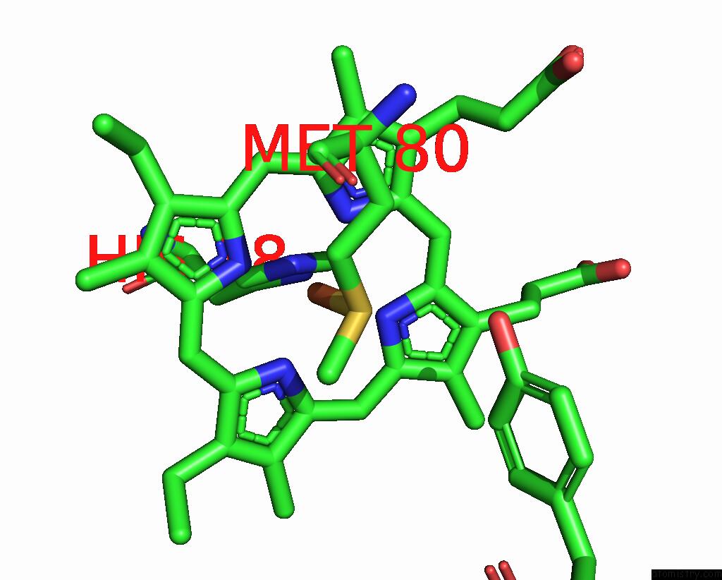
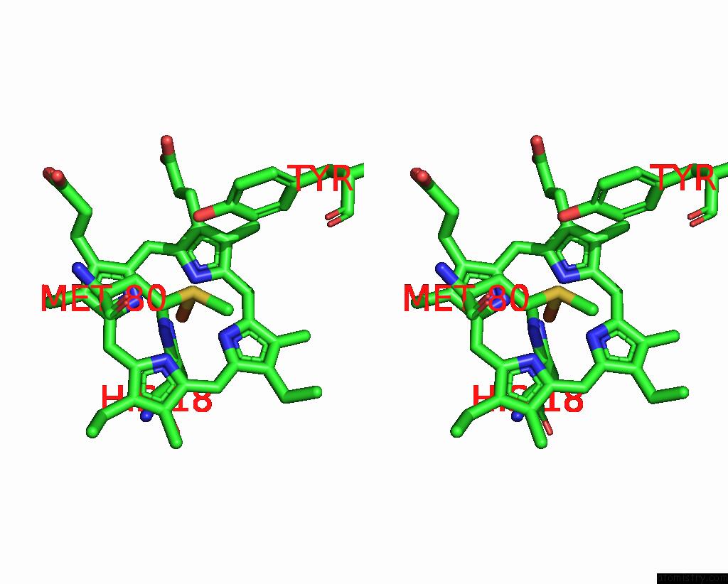
Iron binding site 1 out of 4 in 7bbt
Go back to
Iron binding site 1 out
of 4 in the Structure of Cytochrome C in Complex with A P-Benzyl-Sulfonato- Calix[8]Arene-Peg Pseudorotaxane
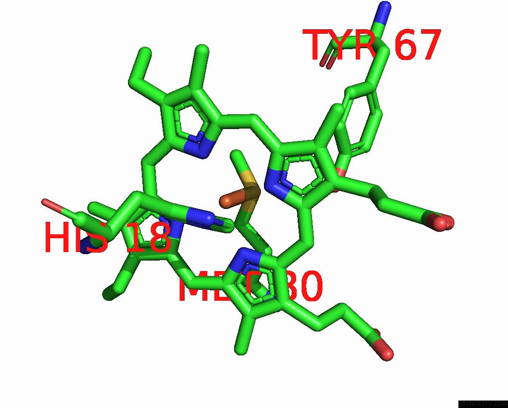
Mono view
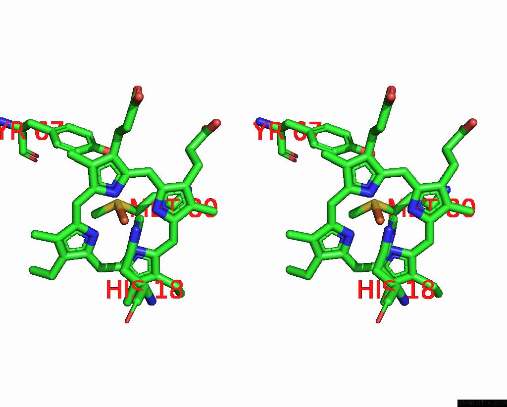
Stereo pair view
Mono view
Stereo pair view
A full contact list of Iron with other atoms in the Fe binding
site number 1 of Structure of Cytochrome C in Complex with A P-Benzyl-Sulfonato- Calix[8]Arene-Peg Pseudorotaxane within 5.0Å range:
|
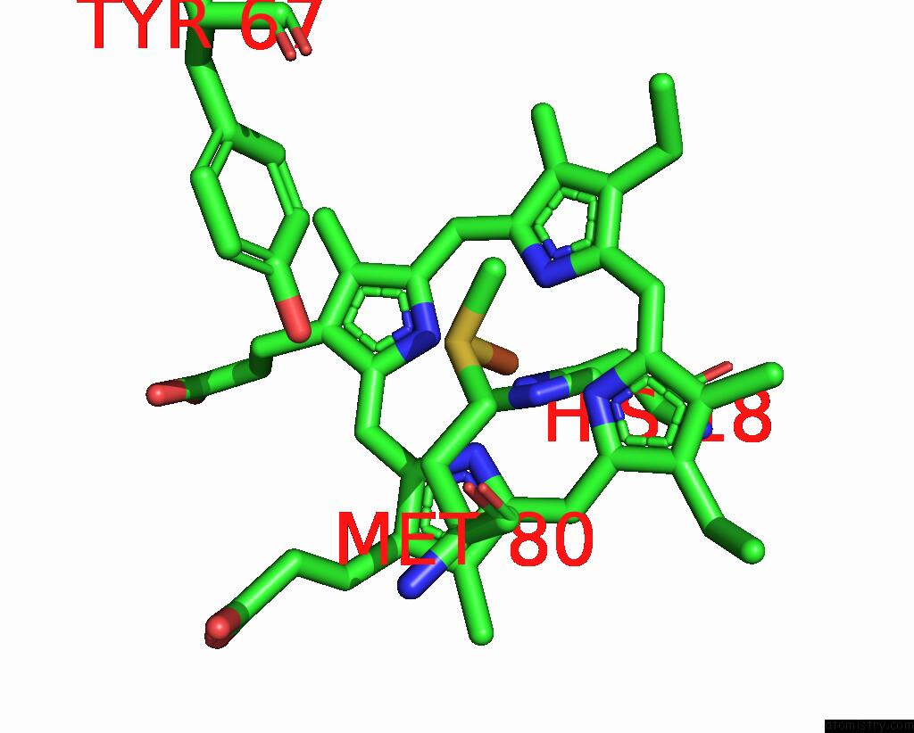
Iron binding site 2 out of 4 in 7bbt
Go back to
Iron binding site 2 out
of 4 in the Structure of Cytochrome C in Complex with A P-Benzyl-Sulfonato- Calix[8]Arene-Peg Pseudorotaxane
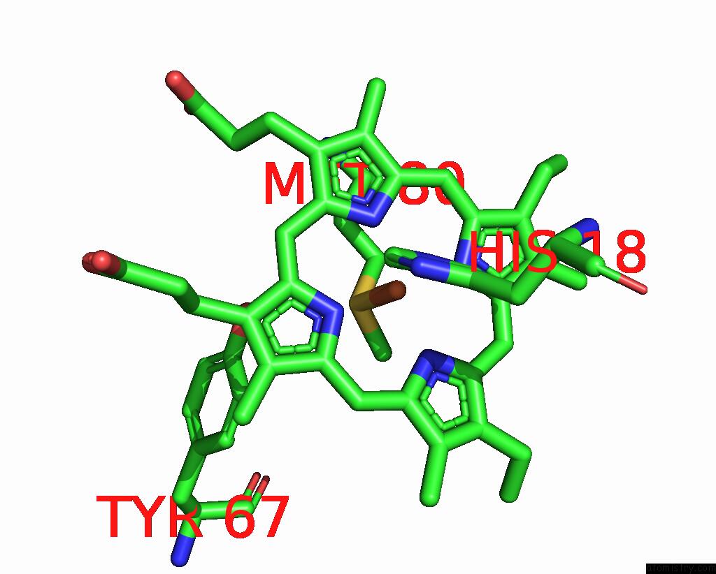
Mono view
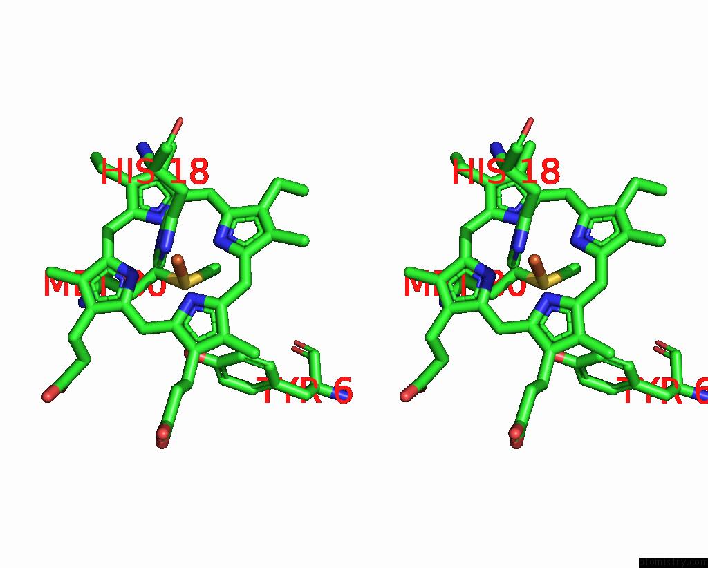
Stereo pair view
Mono view
Stereo pair view
A full contact list of Iron with other atoms in the Fe binding
site number 2 of Structure of Cytochrome C in Complex with A P-Benzyl-Sulfonato- Calix[8]Arene-Peg Pseudorotaxane within 5.0Å range:
|
Iron binding site 3 out of 4 in 7bbt
Go back to
Iron binding site 3 out
of 4 in the Structure of Cytochrome C in Complex with A P-Benzyl-Sulfonato- Calix[8]Arene-Peg Pseudorotaxane
Mono view
Stereo pair view
Mono view
Stereo pair view
A full contact list of Iron with other atoms in the Fe binding
site number 3 of Structure of Cytochrome C in Complex with A P-Benzyl-Sulfonato- Calix[8]Arene-Peg Pseudorotaxane within 5.0Å range:
|
Iron binding site 4 out of 4 in 7bbt
Go back to
Iron binding site 4 out
of 4 in the Structure of Cytochrome C in Complex with A P-Benzyl-Sulfonato- Calix[8]Arene-Peg Pseudorotaxane
Mono view

Stereo pair view
Mono view
Stereo pair view
A full contact list of Iron with other atoms in the Fe binding
site number 4 of Structure of Cytochrome C in Complex with A P-Benzyl-Sulfonato- Calix[8]Arene-Peg Pseudorotaxane within 5.0Å range:
|
Reference:
N.M.Mockler,
K.O.Ramberg,
F.Guagnini,
C.L.Raston,
P.B.Crowley.
Noncovalent Protein-Pseudorotaxane Assembly Incorporating An Extended Arm Calix[8]Arene with Alpha-Helical Recognition Properties. Cryst.Growth Des. V. 21 1424 2021.
ISSN: ISSN 1528-7483
PubMed: 34054353
DOI: 10.1021/ACS.CGD.0C01717
Page generated: Wed Aug 6 20:00:30 2025
ISSN: ISSN 1528-7483
PubMed: 34054353
DOI: 10.1021/ACS.CGD.0C01717
Last articles
Fe in 7JORFe in 7JRD
Fe in 7GEP
Fe in 7JJQ
Fe in 7JJ1
Fe in 7HBI
Fe in 7FJJ
Fe in 7FD1
Fe in 7FDR
Fe in 7FJI