Atomistry » Iron » PDB 7qhf-7r2p » 7qyq
Atomistry »
  Iron »
    PDB 7qhf-7r2p »
      7qyq »

Iron in PDB 7qyq: Crystal Structure of A Dyp-Type Peroxidase From Pseudomonas Putida

Protein crystallography data

The structure of Crystal Structure of A Dyp-Type Peroxidase From Pseudomonas Putida, PDB code: 7qyq was solved by P.T.Borges, D.Silva, C.Frazao, L.O.Martins, with X-Ray Crystallography technique. A brief refinement statistics is given in the table below:

Resolution Low / High (Å) 101.05 / 2.60
Space group P 32 2 1
Cell size a, b, c (Å), α, β, γ (°) 141.951, 141.951, 177.44, 90, 90, 120
R / Rfree (%) 21.9 / 23.5

Iron Binding Sites:

The binding sites of Iron atom in the Crystal Structure of A Dyp-Type Peroxidase From Pseudomonas Putida (pdb code 7qyq). This binding sites where shown within 5.0 Angstroms radius around Iron atom.
In total 4 binding sites of Iron where determined in the Crystal Structure of A Dyp-Type Peroxidase From Pseudomonas Putida, PDB code: 7qyq:
Jump to Iron binding site number: 1; 2; 3; 4;

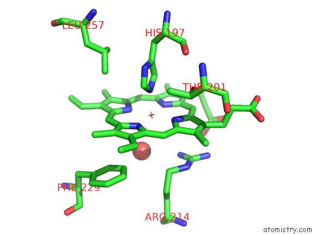
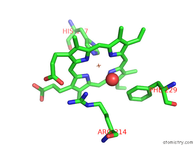
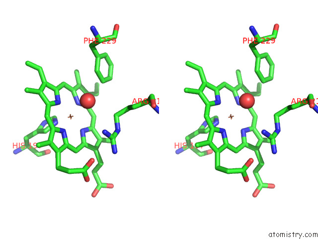
Iron binding site 1 out of 4 in 7qyq

Go back to Iron Binding Sites List in 7qyq
Iron binding site 1 out of 4 in the Crystal Structure of A Dyp-Type Peroxidase From Pseudomonas Putida


Mono view


Stereo pair view

A full contact list of Iron with other atoms in the Fe binding site number 1 of Crystal Structure of A Dyp-Type Peroxidase From Pseudomonas Putida within 5.0Å range:
probe atom residue distance (Å) B Occ
A:Fe301

b:31.4
occ:1.00
FE A:HEM301 0.0 31.4 1.0
NB A:HEM301 2.0 26.9 1.0
ND A:HEM301 2.0 27.4 1.0
NA A:HEM301 2.1 27.0 1.0
NC A:HEM301 2.1 25.3 1.0
NE2 A:HIS197 2.1 53.8 1.0
C1D A:HEM301 3.1 28.7 1.0
C1B A:HEM301 3.1 28.0 1.0
C4B A:HEM301 3.1 29.6 1.0
C4D A:HEM301 3.1 30.0 1.0
C1A A:HEM301 3.1 31.2 1.0
C4C A:HEM301 3.1 27.0 1.0
CE1 A:HIS197 3.1 53.8 1.0
C4A A:HEM301 3.1 28.5 1.0
C1C A:HEM301 3.1 28.6 1.0
CD2 A:HIS197 3.1 53.5 1.0
CHD A:HEM301 3.4 27.4 1.0
CHB A:HEM301 3.4 29.9 1.0
CHA A:HEM301 3.4 31.6 1.0
CHC A:HEM301 3.4 29.5 1.0
O A:HOH440 3.6 35.2 1.0
ND1 A:HIS197 4.2 54.4 1.0
CG A:HIS197 4.3 54.0 1.0
C2D A:HEM301 4.3 32.4 1.0
C3D A:HEM301 4.3 31.8 1.0
C2B A:HEM301 4.3 27.4 1.0
C3B A:HEM301 4.3 30.7 1.0
C2A A:HEM301 4.3 30.1 1.0
C3A A:HEM301 4.3 27.7 1.0
C3C A:HEM301 4.3 28.1 1.0
C2C A:HEM301 4.3 28.7 1.0
NH1 A:ARG214 4.5 39.5 1.0
CD A:ARG214 4.6 35.9 1.0
CE1 A:PHE229 4.7 26.3 1.0
CZ A:ARG214 4.9 38.7 1.0
OG1 A:THR201 4.9 48.7 1.0
NE A:ARG214 4.9 37.1 1.0
CD1 A:LEU257 5.0 31.7 1.0

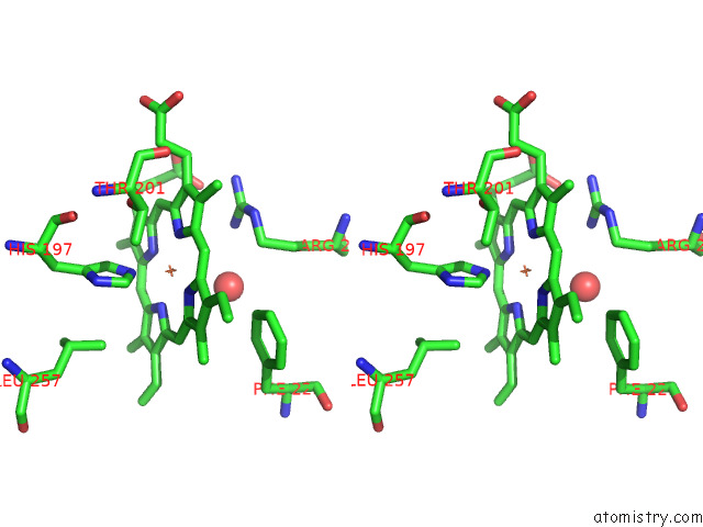
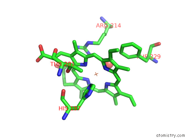
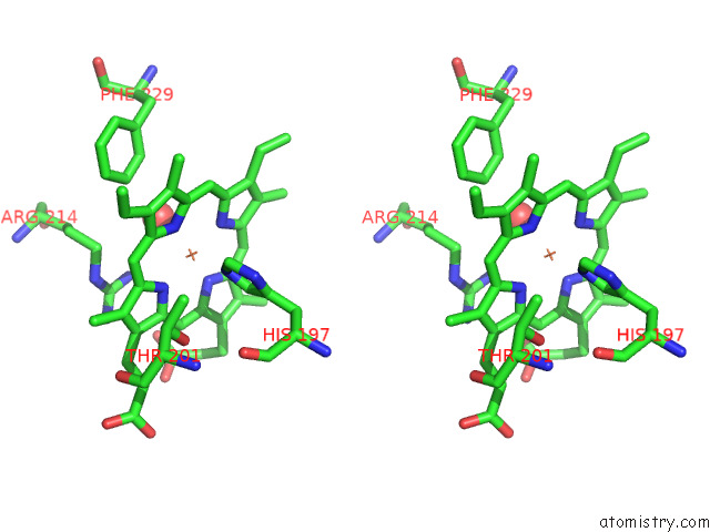
Iron binding site 2 out of 4 in 7qyq

Go back to Iron Binding Sites List in 7qyq
Iron binding site 2 out of 4 in the Crystal Structure of A Dyp-Type Peroxidase From Pseudomonas Putida


Mono view


Stereo pair view

A full contact list of Iron with other atoms in the Fe binding site number 2 of Crystal Structure of A Dyp-Type Peroxidase From Pseudomonas Putida within 5.0Å range:
probe atom residue distance (Å) B Occ
B:Fe301

b:47.4
occ:1.00
FE B:HEM301 0.0 47.4 1.0
ND B:HEM301 2.0 48.1 1.0
NB B:HEM301 2.0 46.8 1.0
NA B:HEM301 2.0 51.4 1.0
NC B:HEM301 2.1 48.3 1.0
NE2 B:HIS197 2.2 52.7 1.0
O B:HOH431 2.9 26.7 1.0
C4B B:HEM301 3.1 48.1 1.0
C1A B:HEM301 3.1 51.1 1.0
C1D B:HEM301 3.1 47.8 1.0
C4D B:HEM301 3.1 48.8 1.0
C4A B:HEM301 3.1 49.5 1.0
C1C B:HEM301 3.1 47.3 1.0
C1B B:HEM301 3.1 44.9 1.0
C4C B:HEM301 3.1 48.1 1.0
CE1 B:HIS197 3.2 51.5 1.0
CD2 B:HIS197 3.2 51.9 1.0
CHC B:HEM301 3.4 48.1 1.0
CHA B:HEM301 3.4 50.1 1.0
CHD B:HEM301 3.4 47.8 1.0
CHB B:HEM301 3.4 46.4 1.0
C2A B:HEM301 4.3 49.9 1.0
C2C B:HEM301 4.3 47.1 1.0
C3A B:HEM301 4.3 50.2 1.0
C3B B:HEM301 4.3 46.6 1.0
C2D B:HEM301 4.3 46.1 1.0
C3C B:HEM301 4.3 47.9 1.0
C2B B:HEM301 4.3 44.2 1.0
C3D B:HEM301 4.3 47.6 1.0
ND1 B:HIS197 4.3 51.5 1.0
CG B:HIS197 4.3 50.7 1.0
NH1 B:ARG214 4.5 44.3 1.0
CD B:ARG214 4.6 38.9 1.0
CE1 B:PHE229 4.7 43.8 1.0
CZ B:ARG214 4.9 43.7 1.0
NE B:ARG214 4.9 41.2 1.0

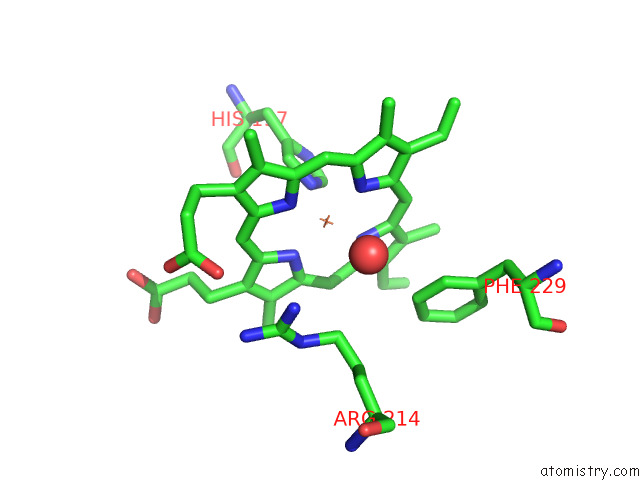
Iron binding site 3 out of 4 in 7qyq

Go back to Iron Binding Sites List in 7qyq
Iron binding site 3 out of 4 in the Crystal Structure of A Dyp-Type Peroxidase From Pseudomonas Putida


Mono view


Stereo pair view

A full contact list of Iron with other atoms in the Fe binding site number 3 of Crystal Structure of A Dyp-Type Peroxidase From Pseudomonas Putida within 5.0Å range:
probe atom residue distance (Å) B Occ
C:Fe301

b:33.5
occ:1.00
FE C:HEM301 0.0 33.5 1.0
NB C:HEM301 2.0 34.0 1.0
ND C:HEM301 2.0 33.8 1.0
NC C:HEM301 2.1 31.6 1.0
NA C:HEM301 2.1 34.7 1.0
NE2 C:HIS197 2.2 55.3 1.0
C1D C:HEM301 3.1 36.1 1.0
C4B C:HEM301 3.1 33.1 1.0
C4C C:HEM301 3.1 33.8 1.0
C1B C:HEM301 3.1 35.7 1.0
C4A C:HEM301 3.1 37.7 1.0
C4D C:HEM301 3.1 36.0 1.0
C1C C:HEM301 3.1 30.7 1.0
C1A C:HEM301 3.1 37.6 1.0
CE1 C:HIS197 3.1 52.8 1.0
CD2 C:HIS197 3.2 54.2 1.0
CHD C:HEM301 3.4 35.4 1.0
CHB C:HEM301 3.4 37.8 1.0
O C:HOH426 3.4 35.3 1.0
CHC C:HEM301 3.4 31.0 1.0
CHA C:HEM301 3.4 37.1 1.0
ND1 C:HIS197 4.3 53.1 1.0
C2D C:HEM301 4.3 37.2 1.0
C3D C:HEM301 4.3 38.8 1.0
C3B C:HEM301 4.3 33.5 1.0
C2B C:HEM301 4.3 33.7 1.0
C3C C:HEM301 4.3 35.1 1.0
C2C C:HEM301 4.3 31.9 1.0
C3A C:HEM301 4.3 40.2 1.0
C2A C:HEM301 4.3 40.7 1.0
CG C:HIS197 4.3 53.3 1.0
CD C:ARG214 4.6 38.0 1.0
NH1 C:ARG214 4.6 39.9 1.0
CE1 C:PHE229 4.8 38.9 1.0
NE C:ARG214 5.0 39.9 1.0
CZ C:ARG214 5.0 40.6 1.0

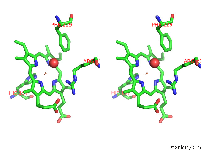
Iron binding site 4 out of 4 in 7qyq

Go back to Iron Binding Sites List in 7qyq
Iron binding site 4 out of 4 in the Crystal Structure of A Dyp-Type Peroxidase From Pseudomonas Putida


Mono view


Stereo pair view

A full contact list of Iron with other atoms in the Fe binding site number 4 of Crystal Structure of A Dyp-Type Peroxidase From Pseudomonas Putida within 5.0Å range:
probe atom residue distance (Å) B Occ
D:Fe301

b:47.4
occ:1.00
FE D:HEM301 0.0 47.4 1.0
NB D:HEM301 2.0 47.9 1.0
ND D:HEM301 2.0 47.6 1.0
NC D:HEM301 2.1 49.2 1.0
NA D:HEM301 2.1 50.2 1.0
NE2 D:HIS197 2.2 33.9 1.0
C4B D:HEM301 3.1 47.5 1.0
C1D D:HEM301 3.1 46.3 1.0
C1B D:HEM301 3.1 48.0 1.0
C4C D:HEM301 3.1 49.9 1.0
C1C D:HEM301 3.1 49.0 1.0
C4A D:HEM301 3.1 50.0 1.0
C4D D:HEM301 3.1 48.0 1.0
C1A D:HEM301 3.1 48.8 1.0
CD2 D:HIS197 3.2 34.5 1.0
CE1 D:HIS197 3.2 34.0 1.0
CHC D:HEM301 3.4 47.2 1.0
CHD D:HEM301 3.4 48.1 1.0
CHB D:HEM301 3.4 50.3 1.0
CHA D:HEM301 3.4 48.4 1.0
O D:HOH425 3.5 27.9 1.0
C3B D:HEM301 4.3 46.3 1.0
C2D D:HEM301 4.3 44.1 1.0
C2B D:HEM301 4.3 45.7 1.0
C3A D:HEM301 4.3 48.2 1.0
C3D D:HEM301 4.3 47.3 1.0
C2A D:HEM301 4.3 47.6 1.0
C2C D:HEM301 4.3 52.5 1.0
C3C D:HEM301 4.3 52.2 1.0
ND1 D:HIS197 4.3 35.1 1.0
NH1 D:ARG214 4.3 37.9 1.0
CG D:HIS197 4.4 35.4 1.0
CD D:ARG214 4.5 36.2 1.0
CE1 D:PHE229 4.6 40.1 1.0
CZ D:ARG214 4.7 39.5 1.0
NE D:ARG214 4.8 40.5 1.0
OG1 D:THR201 5.0 39.1 1.0

Reference:

P.T.Borges, D.Silva, T.F.D.Silva, V.Brissos, M.Canellas, M.F.Lucas, L.Masgrau, E.P.Melo, M.Machuqueiro, C.Frazao, L.O.Martins. Unveiling Molecular Details Behind Improved Activity at Neutral to Alkaline pH of An Engineered Dyp-Type Peroxidase. Comput Struct Biotechnol J V. 20 3899 2022.
ISSN: ESSN 2001-0370
PubMed: 35950185
DOI: 10.1016/J.CSBJ.2022.07.032
Page generated: Wed Apr 5 04:25:59 2023

Last articles

Zn in 8WB0
Zn in 8WAX
Zn in 8WAU
Zn in 8WAZ
Zn in 8WAY
Zn in 8WAV
Zn in 8WAW
Zn in 8WAT
Zn in 8W7M
Zn in 8WD3
© Copyright 2008-2020 by atomistry.com
Home   |    Site Map   |    Copyright   |    Contact us   |    Privacy