Iron »
PDB 7vqg-7w1t »
7vzu »
Iron in PDB 7vzu: The Structure of Gdmn Y82F Mutant
Protein crystallography data
The structure of The Structure of Gdmn Y82F Mutant, PDB code: 7vzu
was solved by
J.Wei,
J.Zheng,
J.Zhou,
Q.Kang,
L.Bai,
with X-Ray Crystallography technique. A brief refinement statistics is given in the table below:
| Resolution Low / High (Å) | 35.83 / 2.30 |
| Space group | P 31 2 1 |
| Cell size a, b, c (Å), α, β, γ (°) | 111.692, 111.692, 231.408, 90, 90, 120 |
| R / Rfree (%) | 18.2 / 22.7 |
Iron Binding Sites:
The binding sites of Iron atom in the The Structure of Gdmn Y82F Mutant
(pdb code 7vzu). This binding sites where shown within
5.0 Angstroms radius around Iron atom.
In total 2 binding sites of Iron where determined in the The Structure of Gdmn Y82F Mutant, PDB code: 7vzu:
Jump to Iron binding site number: 1; 2;
In total 2 binding sites of Iron where determined in the The Structure of Gdmn Y82F Mutant, PDB code: 7vzu:
Jump to Iron binding site number: 1; 2;
Iron binding site 1 out of 2 in 7vzu
Go back to
Iron binding site 1 out
of 2 in the The Structure of Gdmn Y82F Mutant

Mono view
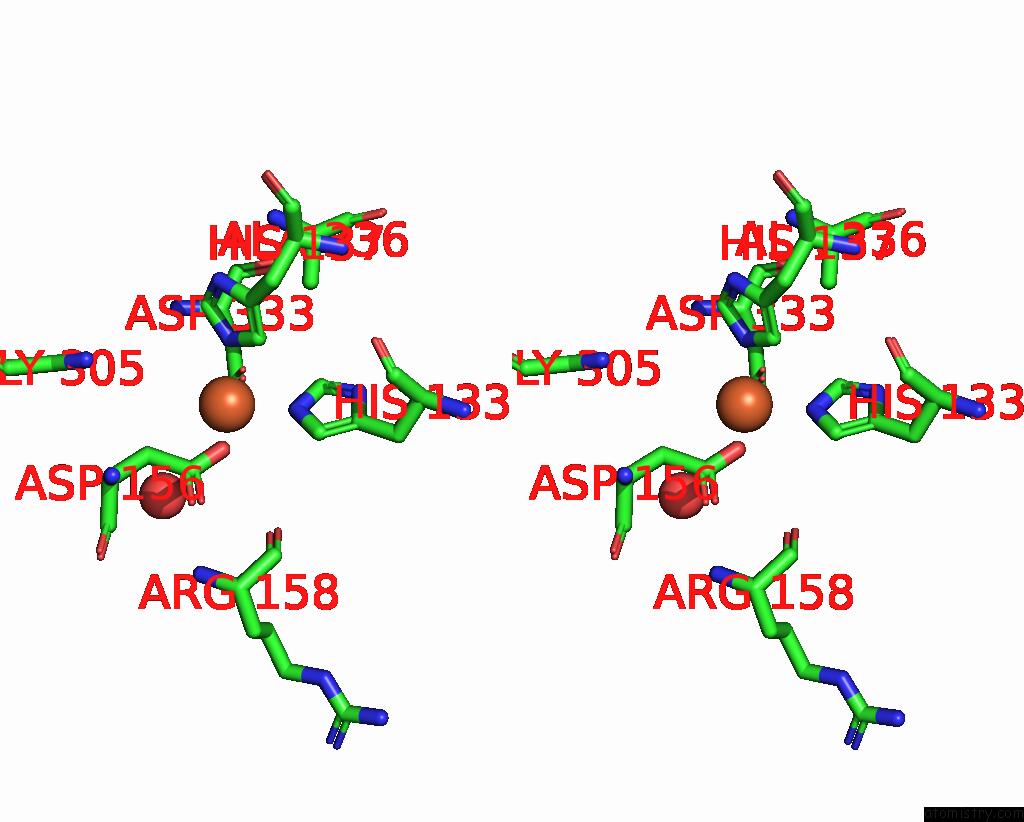
Stereo pair view
Mono view
Stereo pair view
A full contact list of Iron with other atoms in the Fe binding
site number 1 of The Structure of Gdmn Y82F Mutant within 5.0Å range:
|
Iron binding site 2 out of 2 in 7vzu
Go back to
Iron binding site 2 out
of 2 in the The Structure of Gdmn Y82F Mutant

Mono view

Stereo pair view
Mono view
Stereo pair view
A full contact list of Iron with other atoms in the Fe binding
site number 2 of The Structure of Gdmn Y82F Mutant within 5.0Å range:
|
Reference:
J.Wei,
X.Zhang,
Y.Zhou,
X.Cheng,
Z.Lin,
M.Tang,
J.Zheng,
B.Wang,
Q.Kang,
L.Bai.
Endowing Homodimeric Carbamoyltransferase Gdmn with Iterative Functions Through Structural Characterization and Mechanistic Studies. Nat Commun V. 13 6617 2022.
ISSN: ESSN 2041-1723
PubMed: 36329057
DOI: 10.1038/S41467-022-34387-2
Page generated: Thu Aug 7 08:23:50 2025
ISSN: ESSN 2041-1723
PubMed: 36329057
DOI: 10.1038/S41467-022-34387-2
Last articles
Fe in 7Z7SFe in 7Z7R
Fe in 7Z0T
Fe in 7Z0S
Fe in 7Z6Q
Fe in 7Z53
Fe in 7Z2L
Fe in 7Z1U
Fe in 7Z10
Fe in 7YZQ