Iron in PDB 8b4o: Cryo-Em Structure of Cytochrome Bd Oxidase From C. Glutamicum
Iron Binding Sites:
The binding sites of Iron atom in the Cryo-Em Structure of Cytochrome Bd Oxidase From C. Glutamicum
(pdb code 8b4o). This binding sites where shown within
5.0 Angstroms radius around Iron atom.
In total 3 binding sites of Iron where determined in the
Cryo-Em Structure of Cytochrome Bd Oxidase From C. Glutamicum, PDB code: 8b4o:
Jump to Iron binding site number:
1;
2;
3;
Iron binding site 1 out
of 3 in 8b4o
Go back to
Iron Binding Sites List in 8b4o
Iron binding site 1 out
of 3 in the Cryo-Em Structure of Cytochrome Bd Oxidase From C. Glutamicum
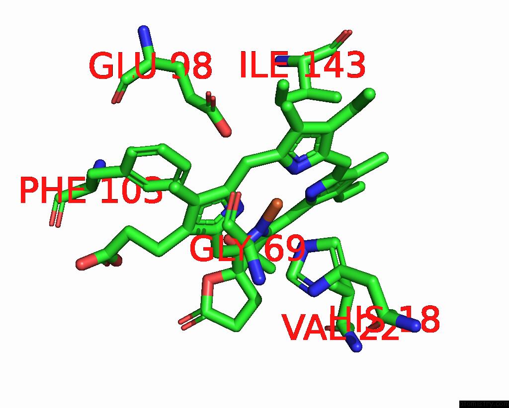
Mono view
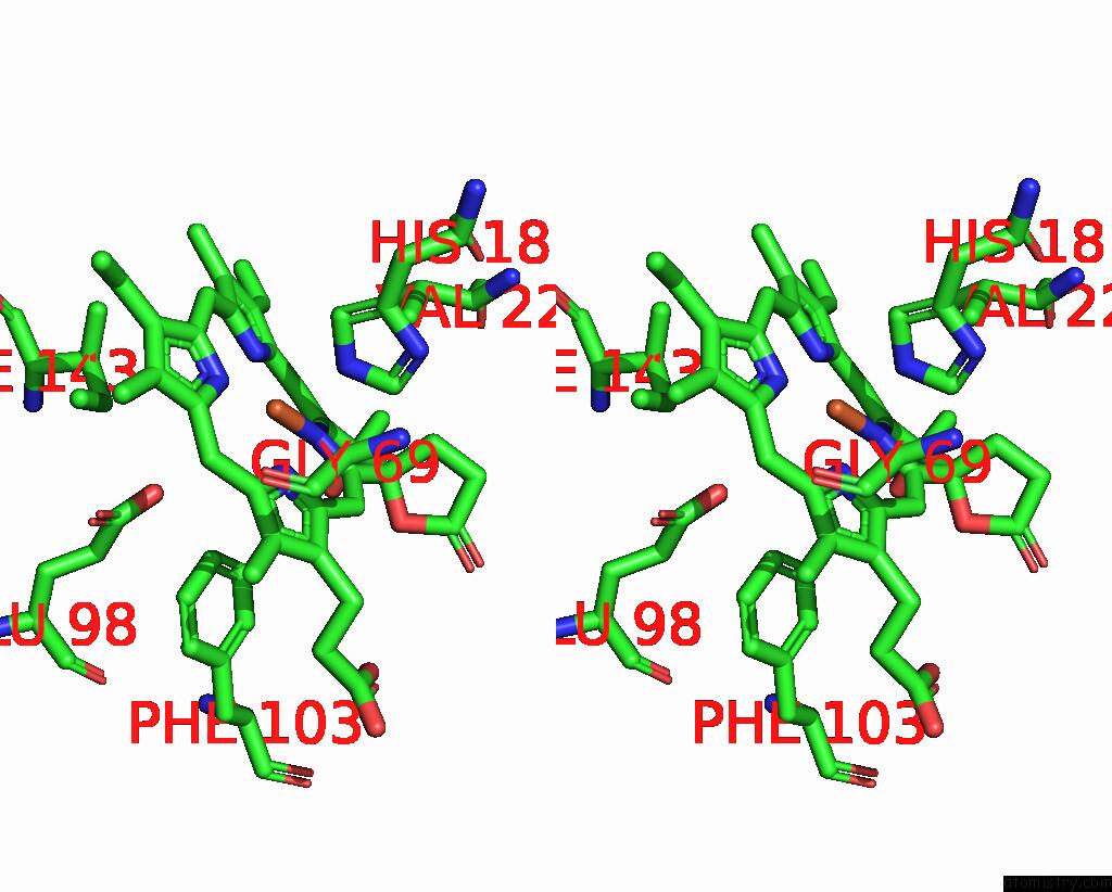
Stereo pair view
|
A full contact list of Iron with other atoms in the Fe binding
site number 1 of Cryo-Em Structure of Cytochrome Bd Oxidase From C. Glutamicum within 5.0Å range:
probe
|
atom
|
residue
|
distance (Å)
|
B
|
Occ
|
A:Fe601
b:16.1
occ:1.00
|
FE
|
A:HDD601
|
0.0
|
16.1
|
1.0
|
ND
|
A:HDD601
|
1.8
|
16.1
|
1.0
|
NA
|
A:HDD601
|
2.1
|
16.1
|
1.0
|
NC
|
A:HDD601
|
2.1
|
16.1
|
1.0
|
NB
|
A:HDD601
|
2.1
|
16.1
|
1.0
|
NE2
|
A:HIS18
|
2.5
|
14.6
|
1.0
|
C4D
|
A:HDD601
|
2.8
|
16.1
|
1.0
|
C1D
|
A:HDD601
|
2.8
|
16.1
|
1.0
|
C4C
|
A:HDD601
|
3.0
|
16.1
|
1.0
|
C1A
|
A:HDD601
|
3.0
|
16.1
|
1.0
|
C1B
|
A:HDD601
|
3.1
|
16.1
|
1.0
|
C4B
|
A:HDD601
|
3.1
|
16.1
|
1.0
|
C4A
|
A:HDD601
|
3.1
|
16.1
|
1.0
|
C1C
|
A:HDD601
|
3.1
|
16.1
|
1.0
|
CHA
|
A:HDD601
|
3.3
|
16.1
|
1.0
|
CHD
|
A:HDD601
|
3.3
|
16.1
|
1.0
|
CHC
|
A:HDD601
|
3.5
|
16.1
|
1.0
|
CHB
|
A:HDD601
|
3.5
|
16.1
|
1.0
|
CD2
|
A:HIS18
|
3.5
|
14.6
|
1.0
|
CE1
|
A:HIS18
|
3.5
|
14.6
|
1.0
|
C3D
|
A:HDD601
|
4.1
|
16.1
|
1.0
|
C2D
|
A:HDD601
|
4.1
|
16.1
|
1.0
|
C3C
|
A:HDD601
|
4.3
|
16.1
|
1.0
|
CZ
|
A:PHE103
|
4.3
|
16.5
|
1.0
|
C2A
|
A:HDD601
|
4.4
|
16.1
|
1.0
|
C2B
|
A:HDD601
|
4.4
|
16.1
|
1.0
|
C3B
|
A:HDD601
|
4.4
|
16.1
|
1.0
|
C3A
|
A:HDD601
|
4.4
|
16.1
|
1.0
|
C2C
|
A:HDD601
|
4.4
|
16.1
|
1.0
|
CG2
|
A:VAL22
|
4.5
|
15.1
|
1.0
|
ND1
|
A:HIS18
|
4.6
|
14.6
|
1.0
|
OE2
|
A:GLU98
|
4.6
|
19.3
|
1.0
|
CG
|
A:HIS18
|
4.6
|
14.6
|
1.0
|
CAD
|
A:HDD601
|
4.7
|
16.1
|
1.0
|
CE1
|
A:PHE103
|
4.7
|
16.5
|
1.0
|
CA
|
A:GLY69
|
4.8
|
16.3
|
1.0
|
CG1
|
A:ILE143
|
4.9
|
18.2
|
1.0
|
OND
|
A:HDD601
|
4.9
|
16.1
|
1.0
|
|
Iron binding site 2 out
of 3 in 8b4o
Go back to
Iron Binding Sites List in 8b4o
Iron binding site 2 out
of 3 in the Cryo-Em Structure of Cytochrome Bd Oxidase From C. Glutamicum
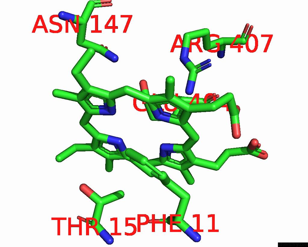
Mono view
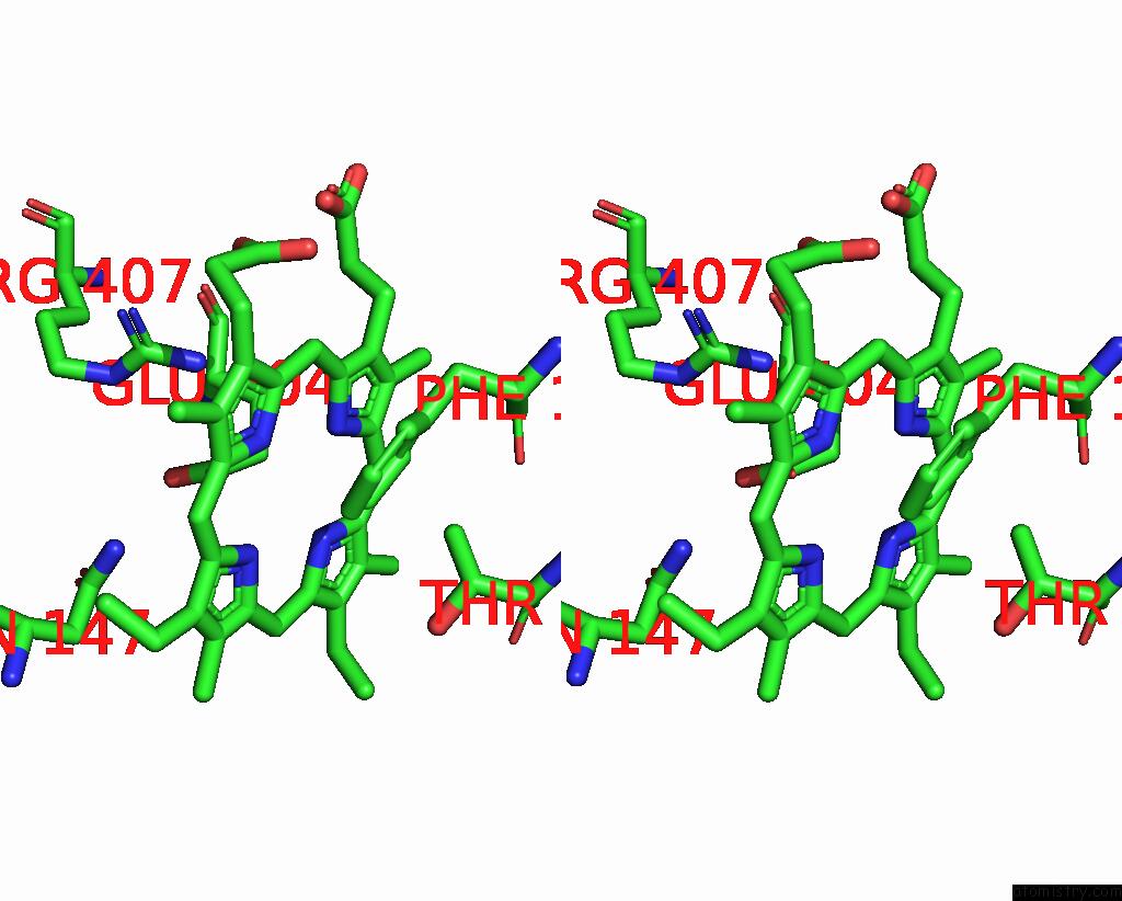
Stereo pair view
|
A full contact list of Iron with other atoms in the Fe binding
site number 2 of Cryo-Em Structure of Cytochrome Bd Oxidase From C. Glutamicum within 5.0Å range:
probe
|
atom
|
residue
|
distance (Å)
|
B
|
Occ
|
A:Fe602
b:25.4
occ:1.00
|
FE
|
A:HEB602
|
0.0
|
25.4
|
1.0
|
NA
|
A:HEB602
|
2.1
|
25.4
|
1.0
|
NB
|
A:HEB602
|
2.2
|
25.4
|
1.0
|
NC
|
A:HEB602
|
2.2
|
25.4
|
1.0
|
ND
|
A:HEB602
|
2.4
|
25.4
|
1.0
|
OE1
|
A:GLU404
|
2.4
|
28.3
|
1.0
|
C1A
|
A:HEB602
|
3.2
|
25.4
|
1.0
|
C1C
|
A:HEB602
|
3.2
|
25.4
|
1.0
|
C1B
|
A:HEB602
|
3.3
|
25.4
|
1.0
|
C4A
|
A:HEB602
|
3.3
|
25.4
|
1.0
|
C4B
|
A:HEB602
|
3.3
|
25.4
|
1.0
|
C4C
|
A:HEB602
|
3.3
|
25.4
|
1.0
|
C4D
|
A:HEB602
|
3.4
|
25.4
|
1.0
|
C1D
|
A:HEB602
|
3.4
|
25.4
|
1.0
|
CD
|
A:GLU404
|
3.4
|
28.3
|
1.0
|
CE1
|
A:PHE11
|
3.6
|
21.4
|
1.0
|
CHC
|
A:HEB602
|
3.7
|
25.4
|
1.0
|
CHB
|
A:HEB602
|
3.7
|
25.4
|
1.0
|
CG
|
A:GLU404
|
3.7
|
28.3
|
1.0
|
CHA
|
A:HEB602
|
3.7
|
25.4
|
1.0
|
CHD
|
A:HEB602
|
3.8
|
25.4
|
1.0
|
CD1
|
A:PHE11
|
3.9
|
21.4
|
1.0
|
NH2
|
A:ARG407
|
4.1
|
29.9
|
1.0
|
C2A
|
A:HEB602
|
4.2
|
25.4
|
1.0
|
C2C
|
A:HEB602
|
4.2
|
25.4
|
1.0
|
C2B
|
A:HEB602
|
4.2
|
25.4
|
1.0
|
CG2
|
A:THR15
|
4.3
|
18.2
|
1.0
|
C3A
|
A:HEB602
|
4.3
|
25.4
|
1.0
|
C3B
|
A:HEB602
|
4.3
|
25.4
|
1.0
|
C3C
|
A:HEB602
|
4.4
|
25.4
|
1.0
|
C2D
|
A:HEB602
|
4.4
|
25.4
|
1.0
|
CB
|
A:GLU404
|
4.5
|
28.3
|
1.0
|
OE2
|
A:GLU404
|
4.5
|
28.3
|
1.0
|
C3D
|
A:HEB602
|
4.6
|
25.4
|
1.0
|
CZ
|
A:PHE11
|
4.6
|
21.4
|
1.0
|
ND2
|
A:ASN147
|
4.9
|
23.6
|
1.0
|
|
Iron binding site 3 out
of 3 in 8b4o
Go back to
Iron Binding Sites List in 8b4o
Iron binding site 3 out
of 3 in the Cryo-Em Structure of Cytochrome Bd Oxidase From C. Glutamicum
Mono view
Stereo pair view
|
A full contact list of Iron with other atoms in the Fe binding
site number 3 of Cryo-Em Structure of Cytochrome Bd Oxidase From C. Glutamicum within 5.0Å range:
probe
|
atom
|
residue
|
distance (Å)
|
B
|
Occ
|
A:Fe603
b:29.6
occ:1.00
|
FE
|
A:HEB603
|
0.0
|
29.6
|
1.0
|
NA
|
A:HEB603
|
2.1
|
29.6
|
1.0
|
NB
|
A:HEB603
|
2.2
|
29.6
|
1.0
|
NC
|
A:HEB603
|
2.2
|
29.6
|
1.0
|
SD
|
A:MET349
|
2.3
|
35.4
|
1.0
|
ND
|
A:HEB603
|
2.4
|
29.6
|
1.0
|
NE2
|
A:HIS185
|
2.4
|
25.0
|
1.0
|
CD2
|
A:HIS185
|
3.1
|
25.0
|
1.0
|
C1A
|
A:HEB603
|
3.2
|
29.6
|
1.0
|
C1C
|
A:HEB603
|
3.2
|
29.6
|
1.0
|
C4A
|
A:HEB603
|
3.3
|
29.6
|
1.0
|
C1B
|
A:HEB603
|
3.3
|
29.6
|
1.0
|
C4B
|
A:HEB603
|
3.3
|
29.6
|
1.0
|
C4C
|
A:HEB603
|
3.3
|
29.6
|
1.0
|
C4D
|
A:HEB603
|
3.4
|
29.6
|
1.0
|
CG
|
A:MET349
|
3.4
|
35.4
|
1.0
|
C1D
|
A:HEB603
|
3.4
|
29.6
|
1.0
|
CE1
|
A:HIS185
|
3.5
|
25.0
|
1.0
|
CHB
|
A:HEB603
|
3.7
|
29.6
|
1.0
|
CHC
|
A:HEB603
|
3.7
|
29.6
|
1.0
|
CHA
|
A:HEB603
|
3.7
|
29.6
|
1.0
|
CHD
|
A:HEB603
|
3.8
|
29.6
|
1.0
|
CE
|
A:MET349
|
3.8
|
35.4
|
1.0
|
C2A
|
A:HEB603
|
4.2
|
29.6
|
1.0
|
C2C
|
A:HEB603
|
4.2
|
29.6
|
1.0
|
C2B
|
A:HEB603
|
4.2
|
29.6
|
1.0
|
C3A
|
A:HEB603
|
4.3
|
29.6
|
1.0
|
C3B
|
A:HEB603
|
4.3
|
29.6
|
1.0
|
CG
|
A:HIS185
|
4.3
|
25.0
|
1.0
|
C3C
|
A:HEB603
|
4.4
|
29.6
|
1.0
|
C2D
|
A:HEB603
|
4.4
|
29.6
|
1.0
|
ND1
|
A:HIS185
|
4.5
|
25.0
|
1.0
|
C3D
|
A:HEB603
|
4.6
|
29.6
|
1.0
|
CB
|
A:MET349
|
4.8
|
35.4
|
1.0
|
|
Reference:
T.N.Grund,
H.Michel,
S.Safarian.
Cryo-Em Structure of Cytochrome Bd Oxidase From C. Glutamicum To Be Published.
Page generated: Fri Aug 9 18:50:26 2024
|