Iron »
PDB 9evv-9fzz »
9fka »
Iron in PDB 9fka: Cryo-Em Structure of the Reduced Cytochrome Bd Oxidase From M. Tuberculosis
Iron Binding Sites:
The binding sites of Iron atom in the Cryo-Em Structure of the Reduced Cytochrome Bd Oxidase From M. Tuberculosis
(pdb code 9fka). This binding sites where shown within
5.0 Angstroms radius around Iron atom.
In total 3 binding sites of Iron where determined in the Cryo-Em Structure of the Reduced Cytochrome Bd Oxidase From M. Tuberculosis, PDB code: 9fka:
Jump to Iron binding site number: 1; 2; 3;
In total 3 binding sites of Iron where determined in the Cryo-Em Structure of the Reduced Cytochrome Bd Oxidase From M. Tuberculosis, PDB code: 9fka:
Jump to Iron binding site number: 1; 2; 3;
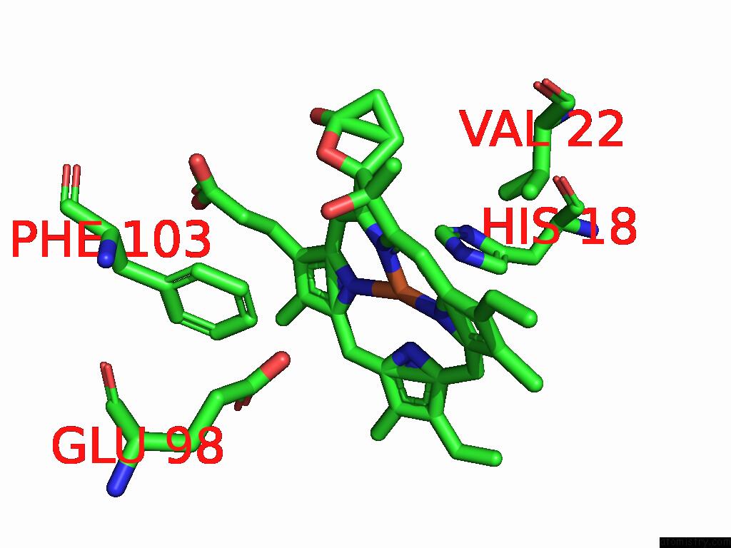
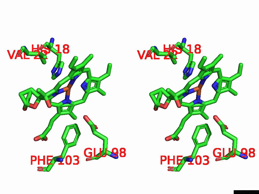
Iron binding site 1 out of 3 in 9fka
Go back to
Iron binding site 1 out
of 3 in the Cryo-Em Structure of the Reduced Cytochrome Bd Oxidase From M. Tuberculosis
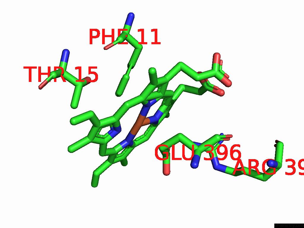
Mono view
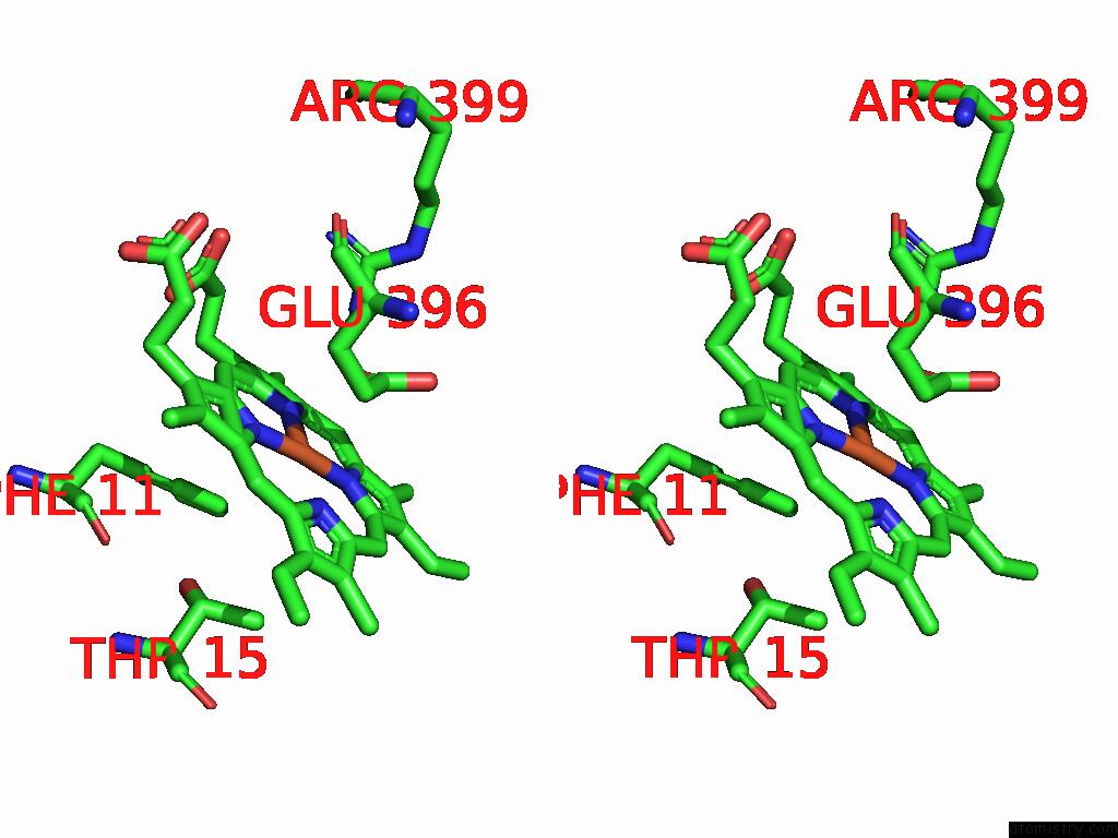
Stereo pair view
Mono view
Stereo pair view
A full contact list of Iron with other atoms in the Fe binding
site number 1 of Cryo-Em Structure of the Reduced Cytochrome Bd Oxidase From M. Tuberculosis within 5.0Å range:
|
Iron binding site 2 out of 3 in 9fka
Go back to
Iron binding site 2 out
of 3 in the Cryo-Em Structure of the Reduced Cytochrome Bd Oxidase From M. Tuberculosis
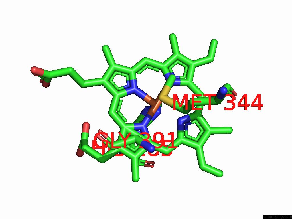
Mono view
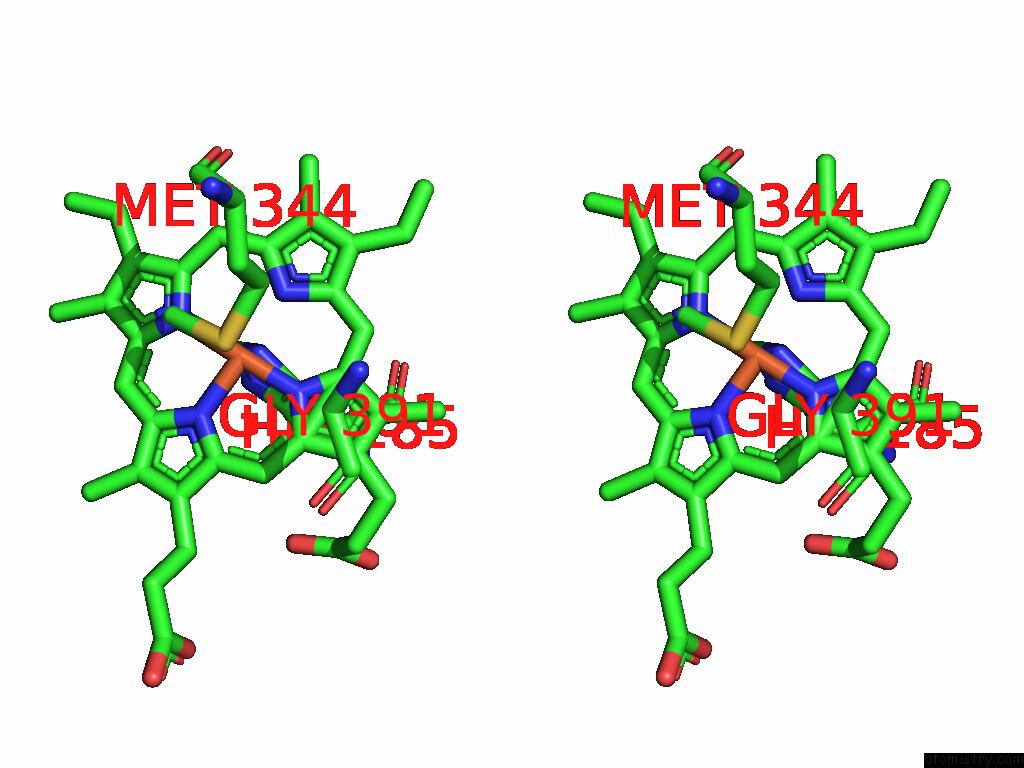
Stereo pair view
Mono view
Stereo pair view
A full contact list of Iron with other atoms in the Fe binding
site number 2 of Cryo-Em Structure of the Reduced Cytochrome Bd Oxidase From M. Tuberculosis within 5.0Å range:
|
Iron binding site 3 out of 3 in 9fka
Go back to
Iron binding site 3 out
of 3 in the Cryo-Em Structure of the Reduced Cytochrome Bd Oxidase From M. Tuberculosis
Mono view
Stereo pair view
Mono view
Stereo pair view
A full contact list of Iron with other atoms in the Fe binding
site number 3 of Cryo-Em Structure of the Reduced Cytochrome Bd Oxidase From M. Tuberculosis within 5.0Å range:
|
Reference:
T.T.Van Der Velden,
K.Kayastha,
C.Y.J.Waterham,
S.Brunle,
L.J.C.Jeuken.
Menaquinone-Specific Turnover By Mycobacterium Tuberculosis Cytochrome Bd Is Redox Regulated By the Q-Loop Disulfide Bond. J.Biol.Chem. V. 301 08094 2024.
ISSN: ESSN 1083-351X
PubMed: 39706268
DOI: 10.1016/J.JBC.2024.108094
Page generated: Tue Feb 25 10:02:22 2025
ISSN: ESSN 1083-351X
PubMed: 39706268
DOI: 10.1016/J.JBC.2024.108094
Last articles
Fe in 2YXOFe in 2YRS
Fe in 2YXC
Fe in 2YNM
Fe in 2YVJ
Fe in 2YP1
Fe in 2YU2
Fe in 2YU1
Fe in 2YQB
Fe in 2YOO