Iron in PDB 3wg7: A 1.9 Angstrom Radiation Damage Free X-Ray Structure of Large (420KDA) Protein By Femtosecond Crystallography
Enzymatic activity of A 1.9 Angstrom Radiation Damage Free X-Ray Structure of Large (420KDA) Protein By Femtosecond Crystallography
All present enzymatic activity of A 1.9 Angstrom Radiation Damage Free X-Ray Structure of Large (420KDA) Protein By Femtosecond Crystallography:
1.9.3.1;
Protein crystallography data
The structure of A 1.9 Angstrom Radiation Damage Free X-Ray Structure of Large (420KDA) Protein By Femtosecond Crystallography, PDB code: 3wg7
was solved by
K.Hirata,
K.Shinzawa-Itoh,
N.Yano,
S.Takemura,
K.Kato,
M.Hatanaka,
K.Muramoto,
T.Kawahara,
T.Tsukihara,
E.Yamashita,
K.Tono,
G.Ueno,
T.Hikima,
H.Murakami,
Y.Inubushi,
M.Yabashi,
T.Ishikawa,
M.Yamamoto,
T.Ogura,
H.Sugimoto,
J.R.Shen,
S.Yoshikawa,
H.Ago,
with X-Ray Crystallography technique. A brief refinement statistics is given in the table below:
Resolution Low / High (Å)
|
40.00 /
1.90
|
Space group
|
P 21 21 21
|
Cell size a, b, c (Å), α, β, γ (°)
|
182.600,
204.510,
178.290,
90.00,
90.00,
90.00
|
R / Rfree (%)
|
19.5 /
23
|
Other elements in 3wg7:
The structure of A 1.9 Angstrom Radiation Damage Free X-Ray Structure of Large (420KDA) Protein By Femtosecond Crystallography also contains other interesting chemical elements:
Iron Binding Sites:
The binding sites of Iron atom in the A 1.9 Angstrom Radiation Damage Free X-Ray Structure of Large (420KDA) Protein By Femtosecond Crystallography
(pdb code 3wg7). This binding sites where shown within
5.0 Angstroms radius around Iron atom.
In total 4 binding sites of Iron where determined in the
A 1.9 Angstrom Radiation Damage Free X-Ray Structure of Large (420KDA) Protein By Femtosecond Crystallography, PDB code: 3wg7:
Jump to Iron binding site number:
1;
2;
3;
4;
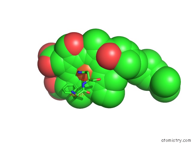
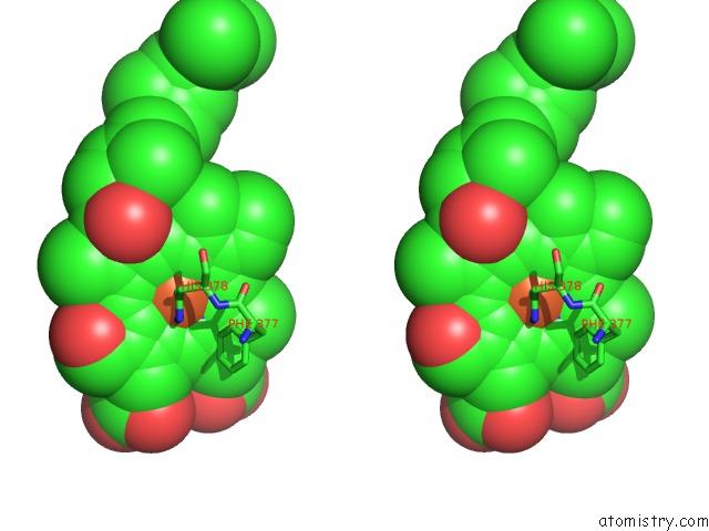
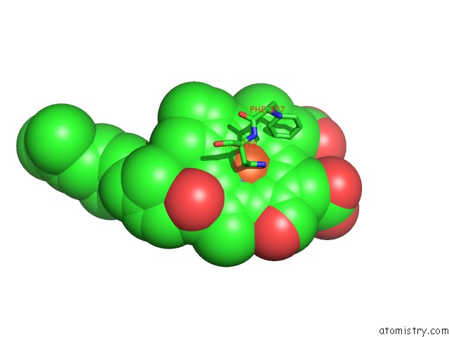
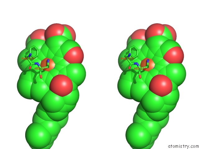
Iron binding site 1 out
of 4 in 3wg7
Go back to
Iron Binding Sites List in 3wg7
Iron binding site 1 out
of 4 in the A 1.9 Angstrom Radiation Damage Free X-Ray Structure of Large (420KDA) Protein By Femtosecond Crystallography
 Mono view
 Stereo pair view
|
A full contact list of Iron with other atoms in the Fe binding
site number 1 of A 1.9 Angstrom Radiation Damage Free X-Ray Structure of Large (420KDA) Protein By Femtosecond Crystallography within 5.0Å range:
probe
|
atom
|
residue
|
distance (Å)
|
B
|
Occ
|
A:Fe601
b:15.5
occ:1.00
|
FE
|
A:HEA601
|
0.0
|
15.5
|
1.0
|
NE2
|
A:HIS61
|
2.0
|
18.5
|
1.0
|
ND
|
A:HEA601
|
2.1
|
13.6
|
1.0
|
NB
|
A:HEA601
|
2.1
|
12.7
|
1.0
|
NC
|
A:HEA601
|
2.2
|
12.1
|
1.0
|
NE2
|
A:HIS378
|
2.2
|
17.9
|
1.0
|
NA
|
A:HEA601
|
2.3
|
13.1
|
1.0
|
CE1
|
A:HIS61
|
3.0
|
15.4
|
1.0
|
CD2
|
A:HIS61
|
3.0
|
17.9
|
1.0
|
CD2
|
A:HIS378
|
3.0
|
13.5
|
1.0
|
C1D
|
A:HEA601
|
3.0
|
12.5
|
1.0
|
C4D
|
A:HEA601
|
3.1
|
13.1
|
1.0
|
C4B
|
A:HEA601
|
3.1
|
12.9
|
1.0
|
C4C
|
A:HEA601
|
3.1
|
12.3
|
1.0
|
C1B
|
A:HEA601
|
3.1
|
13.1
|
1.0
|
C1C
|
A:HEA601
|
3.1
|
11.9
|
1.0
|
C4A
|
A:HEA601
|
3.2
|
11.4
|
1.0
|
C1A
|
A:HEA601
|
3.2
|
11.9
|
1.0
|
CE1
|
A:HIS378
|
3.2
|
14.3
|
1.0
|
CHB
|
A:HEA601
|
3.4
|
12.0
|
1.0
|
CHD
|
A:HEA601
|
3.4
|
11.0
|
1.0
|
CHC
|
A:HEA601
|
3.4
|
12.6
|
1.0
|
CHA
|
A:HEA601
|
3.5
|
12.5
|
1.0
|
ND1
|
A:HIS61
|
4.1
|
14.1
|
1.0
|
CG
|
A:HIS61
|
4.1
|
15.2
|
1.0
|
CG
|
A:HIS378
|
4.2
|
13.4
|
1.0
|
C2D
|
A:HEA601
|
4.3
|
14.2
|
1.0
|
C3D
|
A:HEA601
|
4.3
|
13.9
|
1.0
|
ND1
|
A:HIS378
|
4.3
|
14.5
|
1.0
|
C2C
|
A:HEA601
|
4.3
|
12.4
|
1.0
|
C3C
|
A:HEA601
|
4.3
|
12.8
|
1.0
|
C3B
|
A:HEA601
|
4.3
|
12.7
|
1.0
|
C2B
|
A:HEA601
|
4.4
|
12.3
|
1.0
|
C2A
|
A:HEA601
|
4.4
|
12.2
|
1.0
|
C3A
|
A:HEA601
|
4.5
|
11.7
|
1.0
|
CE2
|
A:PHE377
|
4.8
|
11.5
|
1.0
|
|
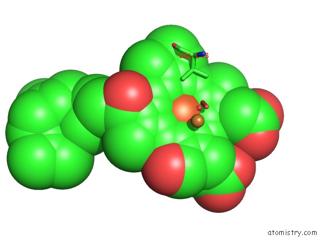
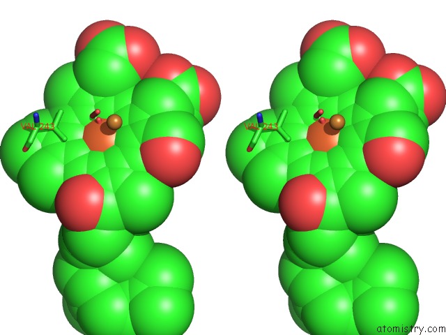
Iron binding site 2 out
of 4 in 3wg7
Go back to
Iron Binding Sites List in 3wg7
Iron binding site 2 out
of 4 in the A 1.9 Angstrom Radiation Damage Free X-Ray Structure of Large (420KDA) Protein By Femtosecond Crystallography
 Mono view
 Stereo pair view
|
A full contact list of Iron with other atoms in the Fe binding
site number 2 of A 1.9 Angstrom Radiation Damage Free X-Ray Structure of Large (420KDA) Protein By Femtosecond Crystallography within 5.0Å range:
probe
|
atom
|
residue
|
distance (Å)
|
B
|
Occ
|
A:Fe602
b:14.8
occ:1.00
|
FE
|
A:HEA602
|
0.0
|
14.8
|
1.0
|
ND
|
A:HEA602
|
2.0
|
11.0
|
1.0
|
NB
|
A:HEA602
|
2.0
|
11.7
|
1.0
|
NE2
|
A:HIS376
|
2.1
|
13.3
|
1.0
|
NC
|
A:HEA602
|
2.1
|
13.0
|
1.0
|
NA
|
A:HEA602
|
2.2
|
11.2
|
1.0
|
O1
|
A:PER606
|
2.4
|
10.2
|
0.9
|
C4D
|
A:HEA602
|
3.0
|
10.6
|
1.0
|
C4B
|
A:HEA602
|
3.1
|
12.7
|
1.0
|
CD2
|
A:HIS376
|
3.1
|
13.1
|
1.0
|
CE1
|
A:HIS376
|
3.1
|
15.2
|
1.0
|
C4C
|
A:HEA602
|
3.1
|
11.7
|
1.0
|
C4A
|
A:HEA602
|
3.1
|
12.2
|
1.0
|
C1B
|
A:HEA602
|
3.1
|
12.8
|
1.0
|
C1D
|
A:HEA602
|
3.1
|
10.4
|
1.0
|
C1C
|
A:HEA602
|
3.1
|
11.7
|
1.0
|
C1A
|
A:HEA602
|
3.1
|
11.8
|
1.0
|
CHB
|
A:HEA602
|
3.4
|
11.6
|
1.0
|
CHA
|
A:HEA602
|
3.5
|
11.2
|
1.0
|
CHC
|
A:HEA602
|
3.5
|
12.4
|
1.0
|
CHD
|
A:HEA602
|
3.5
|
11.4
|
1.0
|
O2
|
A:PER606
|
3.8
|
12.7
|
0.9
|
O1
|
A:PER606
|
3.8
|
11.7
|
0.1
|
ND1
|
A:HIS376
|
4.2
|
15.0
|
1.0
|
CG
|
A:HIS376
|
4.2
|
13.5
|
1.0
|
C3D
|
A:HEA602
|
4.3
|
11.1
|
1.0
|
C3B
|
A:HEA602
|
4.3
|
12.9
|
1.0
|
C3C
|
A:HEA602
|
4.3
|
12.3
|
1.0
|
C2C
|
A:HEA602
|
4.3
|
13.5
|
1.0
|
C2B
|
A:HEA602
|
4.3
|
12.4
|
1.0
|
C2D
|
A:HEA602
|
4.3
|
10.2
|
1.0
|
C2A
|
A:HEA602
|
4.3
|
14.0
|
1.0
|
C3A
|
A:HEA602
|
4.4
|
12.5
|
1.0
|
O2
|
A:PER606
|
4.8
|
11.2
|
0.1
|
CU
|
A:CU603
|
4.9
|
15.8
|
1.0
|
CA
|
A:GLY355
|
5.0
|
11.4
|
1.0
|
|
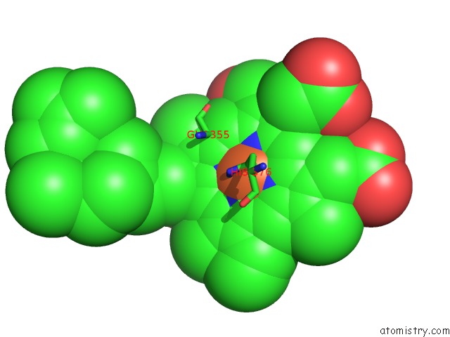
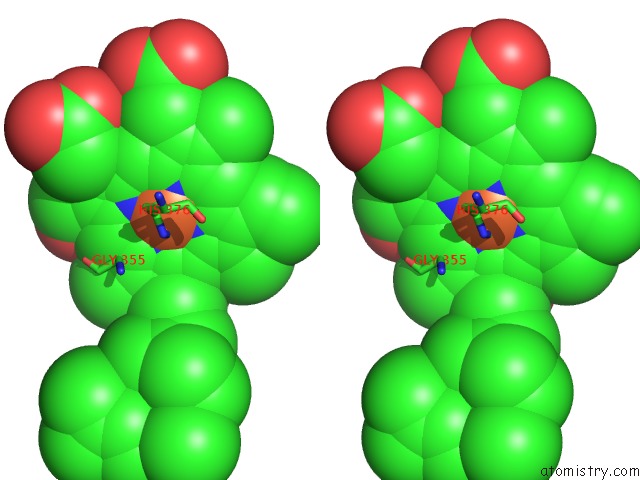
Iron binding site 3 out
of 4 in 3wg7
Go back to
Iron Binding Sites List in 3wg7
Iron binding site 3 out
of 4 in the A 1.9 Angstrom Radiation Damage Free X-Ray Structure of Large (420KDA) Protein By Femtosecond Crystallography
 Mono view
 Stereo pair view
|
A full contact list of Iron with other atoms in the Fe binding
site number 3 of A 1.9 Angstrom Radiation Damage Free X-Ray Structure of Large (420KDA) Protein By Femtosecond Crystallography within 5.0Å range:
probe
|
atom
|
residue
|
distance (Å)
|
B
|
Occ
|
N:Fe601
b:13.9
occ:1.00
|
FE
|
N:HEA601
|
0.0
|
13.9
|
1.0
|
NB
|
N:HEA601
|
2.0
|
12.3
|
1.0
|
NE2
|
N:HIS378
|
2.0
|
12.3
|
1.0
|
NE2
|
N:HIS61
|
2.0
|
12.2
|
1.0
|
ND
|
N:HEA601
|
2.1
|
13.1
|
1.0
|
NA
|
N:HEA601
|
2.1
|
14.8
|
1.0
|
NC
|
N:HEA601
|
2.2
|
13.6
|
1.0
|
CD2
|
N:HIS378
|
3.0
|
13.2
|
1.0
|
CE1
|
N:HIS378
|
3.0
|
14.8
|
1.0
|
CD2
|
N:HIS61
|
3.0
|
13.8
|
1.0
|
C4B
|
N:HEA601
|
3.0
|
12.2
|
1.0
|
C1B
|
N:HEA601
|
3.0
|
12.8
|
1.0
|
CE1
|
N:HIS61
|
3.1
|
12.5
|
1.0
|
C4D
|
N:HEA601
|
3.1
|
12.4
|
1.0
|
C1D
|
N:HEA601
|
3.1
|
15.4
|
1.0
|
C4A
|
N:HEA601
|
3.1
|
12.7
|
1.0
|
C1A
|
N:HEA601
|
3.1
|
12.7
|
1.0
|
C4C
|
N:HEA601
|
3.1
|
13.9
|
1.0
|
C1C
|
N:HEA601
|
3.2
|
12.5
|
1.0
|
CHA
|
N:HEA601
|
3.4
|
13.3
|
1.0
|
CHB
|
N:HEA601
|
3.5
|
12.4
|
1.0
|
CHD
|
N:HEA601
|
3.5
|
14.5
|
1.0
|
CHC
|
N:HEA601
|
3.5
|
11.1
|
1.0
|
ND1
|
N:HIS378
|
4.1
|
12.9
|
1.0
|
CG
|
N:HIS378
|
4.1
|
12.7
|
1.0
|
ND1
|
N:HIS61
|
4.2
|
13.4
|
1.0
|
CG
|
N:HIS61
|
4.2
|
13.0
|
1.0
|
C2B
|
N:HEA601
|
4.2
|
10.7
|
1.0
|
C3B
|
N:HEA601
|
4.2
|
11.3
|
1.0
|
C3A
|
N:HEA601
|
4.3
|
14.1
|
1.0
|
C2D
|
N:HEA601
|
4.3
|
14.4
|
1.0
|
C3D
|
N:HEA601
|
4.3
|
13.0
|
1.0
|
C2A
|
N:HEA601
|
4.3
|
12.6
|
1.0
|
C3C
|
N:HEA601
|
4.4
|
12.8
|
1.0
|
C2C
|
N:HEA601
|
4.4
|
12.5
|
1.0
|
CE2
|
N:PHE377
|
4.9
|
11.7
|
1.0
|
|
Iron binding site 4 out
of 4 in 3wg7
Go back to
Iron Binding Sites List in 3wg7
Iron binding site 4 out
of 4 in the A 1.9 Angstrom Radiation Damage Free X-Ray Structure of Large (420KDA) Protein By Femtosecond Crystallography
 Mono view
 Stereo pair view
|
A full contact list of Iron with other atoms in the Fe binding
site number 4 of A 1.9 Angstrom Radiation Damage Free X-Ray Structure of Large (420KDA) Protein By Femtosecond Crystallography within 5.0Å range:
probe
|
atom
|
residue
|
distance (Å)
|
B
|
Occ
|
N:Fe602
b:14.1
occ:1.00
|
FE
|
N:HEA602
|
0.0
|
14.1
|
1.0
|
NB
|
N:HEA602
|
2.0
|
13.3
|
1.0
|
ND
|
N:HEA602
|
2.0
|
11.2
|
1.0
|
NA
|
N:HEA602
|
2.1
|
12.3
|
1.0
|
NC
|
N:HEA602
|
2.1
|
11.9
|
1.0
|
NE2
|
N:HIS376
|
2.2
|
11.1
|
1.0
|
O1
|
N:PER606
|
2.4
|
10.2
|
0.9
|
C1B
|
N:HEA602
|
3.0
|
11.3
|
1.0
|
C4D
|
N:HEA602
|
3.0
|
11.0
|
1.0
|
C1D
|
N:HEA602
|
3.0
|
12.5
|
1.0
|
C1A
|
N:HEA602
|
3.1
|
11.0
|
1.0
|
C4B
|
N:HEA602
|
3.1
|
12.9
|
1.0
|
C4A
|
N:HEA602
|
3.1
|
11.6
|
1.0
|
C4C
|
N:HEA602
|
3.1
|
12.7
|
1.0
|
C1C
|
N:HEA602
|
3.1
|
12.7
|
1.0
|
CD2
|
N:HIS376
|
3.1
|
12.0
|
1.0
|
CE1
|
N:HIS376
|
3.2
|
11.2
|
1.0
|
CHB
|
N:HEA602
|
3.4
|
11.4
|
1.0
|
CHD
|
N:HEA602
|
3.5
|
11.6
|
1.0
|
CHA
|
N:HEA602
|
3.5
|
10.5
|
1.0
|
CHC
|
N:HEA602
|
3.5
|
12.6
|
1.0
|
O1
|
N:PER606
|
3.7
|
11.7
|
0.1
|
O2
|
N:PER606
|
3.8
|
12.7
|
0.9
|
C2B
|
N:HEA602
|
4.2
|
10.7
|
1.0
|
C3B
|
N:HEA602
|
4.2
|
11.3
|
1.0
|
C2D
|
N:HEA602
|
4.2
|
11.3
|
1.0
|
C3D
|
N:HEA602
|
4.2
|
11.2
|
1.0
|
C2A
|
N:HEA602
|
4.2
|
11.3
|
1.0
|
C3A
|
N:HEA602
|
4.3
|
11.3
|
1.0
|
ND1
|
N:HIS376
|
4.3
|
12.1
|
1.0
|
CG
|
N:HIS376
|
4.3
|
11.2
|
1.0
|
C2C
|
N:HEA602
|
4.3
|
13.0
|
1.0
|
C3C
|
N:HEA602
|
4.4
|
13.8
|
1.0
|
O2
|
N:PER606
|
4.7
|
11.2
|
0.1
|
CG2
|
N:VAL243
|
4.9
|
9.8
|
1.0
|
CU
|
N:CU603
|
4.9
|
14.7
|
1.0
|
|
Reference:
K.Hirata,
K.Shinzawa-Itoh,
N.Yano,
S.Takemura,
K.Kato,
M.Hatanaka,
K.Muramoto,
T.Kawahara,
T.Tsukihara,
E.Yamashita,
K.Tono,
G.Ueno,
T.Hikima,
H.Murakami,
Y.Inubushi,
M.Yabashi,
T.Ishikawa,
M.Yamamoto,
T.Ogura,
H.Sugimoto,
J.R.Shen,
S.Yoshikawa,
H.Ago.
Determination of Damage-Free Crystal Structure of An X-Ray-Sensitive Protein Using An Xfel. Nat.Methods 2014.
ISSN: ESSN 1548-7105
PubMed: 24813624
DOI: 10.1038/NMETH.2962
Page generated: Sun Aug 4 22:39:57 2024
|