Iron »
PDB 4ryx-4toe »
4s1c »
Iron in PDB 4s1c: Crystal Structure of L. Monocytogenes Phosphodiesterase Pgph Hd Domain
Protein crystallography data
The structure of Crystal Structure of L. Monocytogenes Phosphodiesterase Pgph Hd Domain, PDB code: 4s1c
was solved by
S.Luo,
L.Tong,
with X-Ray Crystallography technique. A brief refinement statistics is given in the table below:
| Resolution Low / High (Å) | 24.60 / 2.40 |
| Space group | P 61 |
| Cell size a, b, c (Å), α, β, γ (°) | 116.562, 116.562, 91.783, 90.00, 90.00, 120.00 |
| R / Rfree (%) | 18.5 / 23.1 |
Iron Binding Sites:
The binding sites of Iron atom in the Crystal Structure of L. Monocytogenes Phosphodiesterase Pgph Hd Domain
(pdb code 4s1c). This binding sites where shown within
5.0 Angstroms radius around Iron atom.
In total 4 binding sites of Iron where determined in the Crystal Structure of L. Monocytogenes Phosphodiesterase Pgph Hd Domain, PDB code: 4s1c:
Jump to Iron binding site number: 1; 2; 3; 4;
In total 4 binding sites of Iron where determined in the Crystal Structure of L. Monocytogenes Phosphodiesterase Pgph Hd Domain, PDB code: 4s1c:
Jump to Iron binding site number: 1; 2; 3; 4;
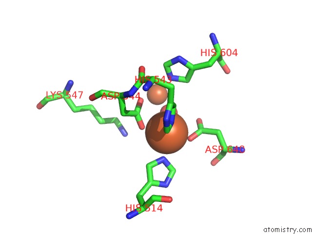
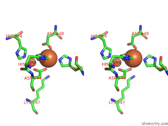
Iron binding site 1 out of 4 in 4s1c
Go back to
Iron binding site 1 out
of 4 in the Crystal Structure of L. Monocytogenes Phosphodiesterase Pgph Hd Domain
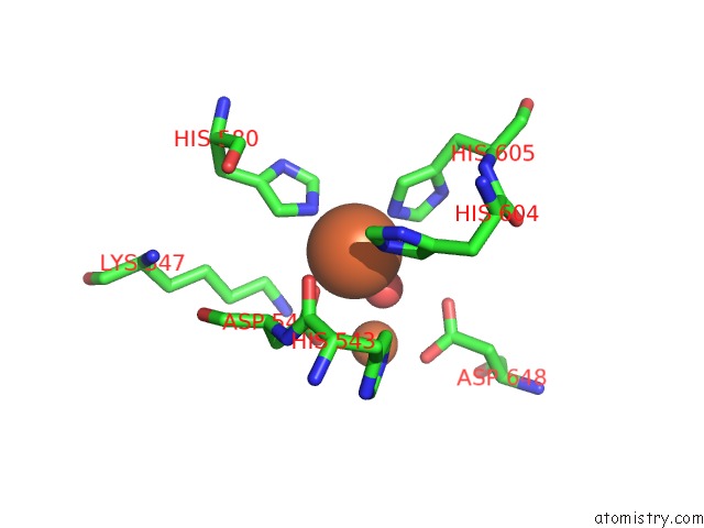
Mono view
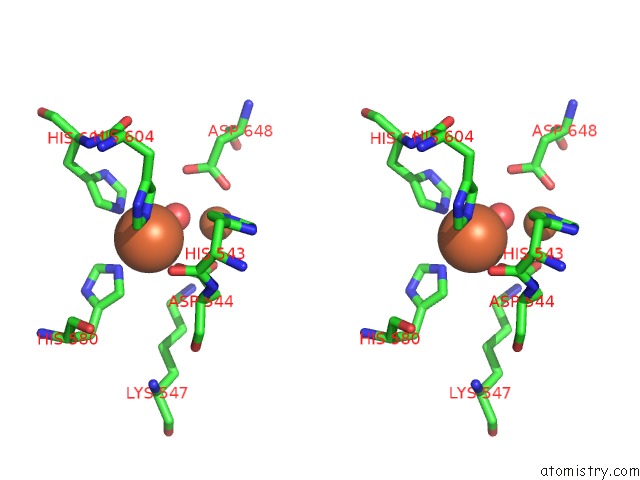
Stereo pair view
Mono view
Stereo pair view
A full contact list of Iron with other atoms in the Fe binding
site number 1 of Crystal Structure of L. Monocytogenes Phosphodiesterase Pgph Hd Domain within 5.0Å range:
|
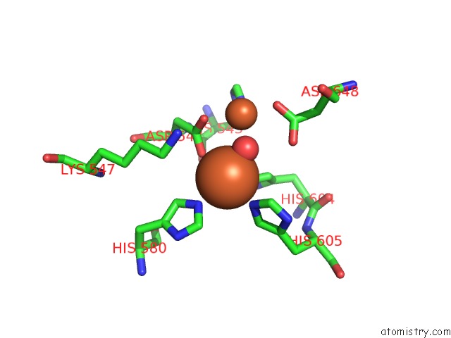
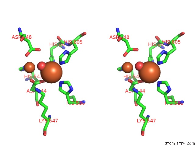
Iron binding site 2 out of 4 in 4s1c
Go back to
Iron binding site 2 out
of 4 in the Crystal Structure of L. Monocytogenes Phosphodiesterase Pgph Hd Domain
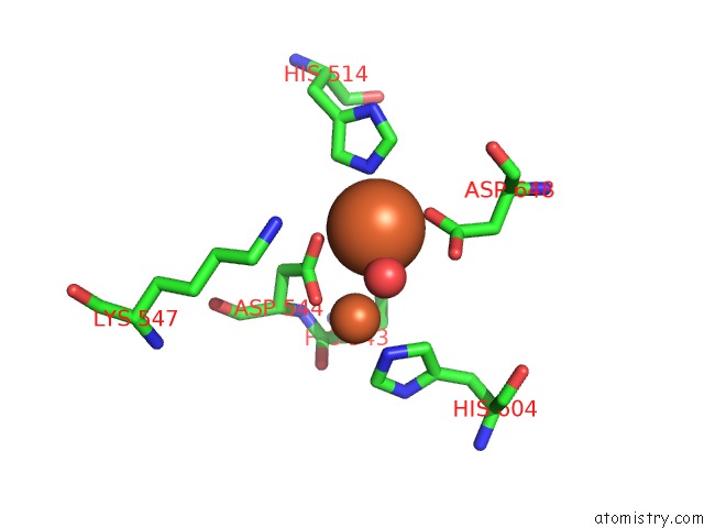
Mono view
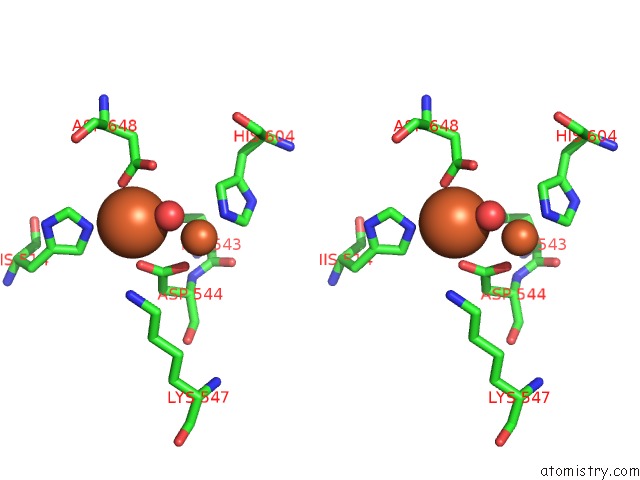
Stereo pair view
Mono view
Stereo pair view
A full contact list of Iron with other atoms in the Fe binding
site number 2 of Crystal Structure of L. Monocytogenes Phosphodiesterase Pgph Hd Domain within 5.0Å range:
|
Iron binding site 3 out of 4 in 4s1c
Go back to
Iron binding site 3 out
of 4 in the Crystal Structure of L. Monocytogenes Phosphodiesterase Pgph Hd Domain
Mono view
Stereo pair view
Mono view
Stereo pair view
A full contact list of Iron with other atoms in the Fe binding
site number 3 of Crystal Structure of L. Monocytogenes Phosphodiesterase Pgph Hd Domain within 5.0Å range:
|
Iron binding site 4 out of 4 in 4s1c
Go back to
Iron binding site 4 out
of 4 in the Crystal Structure of L. Monocytogenes Phosphodiesterase Pgph Hd Domain
Mono view
Stereo pair view
Mono view
Stereo pair view
A full contact list of Iron with other atoms in the Fe binding
site number 4 of Crystal Structure of L. Monocytogenes Phosphodiesterase Pgph Hd Domain within 5.0Å range:
|
Reference:
T.N.Huynh,
S.Luo,
D.Pensinger,
J.D.Sauer,
L.Tong,
J.J.Woodward.
An Hd-Domain Phosphodiesterase Mediates Cooperative Hydrolysis of C-Di-Amp to Affect Bacterial Growth and Virulence. Proc.Natl.Acad.Sci.Usa 2015.
ISSN: ESSN 1091-6490
PubMed: 25583510
DOI: 10.1073/PNAS.1416485112
Page generated: Tue Aug 5 14:28:38 2025
ISSN: ESSN 1091-6490
PubMed: 25583510
DOI: 10.1073/PNAS.1416485112
Last articles
Fe in 6CYSFe in 6CXV
Fe in 6CXU
Fe in 6CSB
Fe in 6CWW
Fe in 6CVC
Fe in 6CUN
Fe in 6CTC
Fe in 6CSD
Fe in 6CUK