Iron »
PDB 4wha-4x33 »
4wq0 »
Iron in PDB 4wq0: Crystal Structure of Cytochrome P450 CYP107W1 From Streptomyces Avermitilis in Complex with Oligomycin A
Protein crystallography data
The structure of Crystal Structure of Cytochrome P450 CYP107W1 From Streptomyces Avermitilis in Complex with Oligomycin A, PDB code: 4wq0
was solved by
L.W.Kang,
D.H.Kim,
T.V.Pham,
S.H.Han,
with X-Ray Crystallography technique. A brief refinement statistics is given in the table below:
| Resolution Low / High (Å) | 40.42 / 2.70 |
| Space group | P 43 21 2 |
| Cell size a, b, c (Å), α, β, γ (°) | 127.812, 127.812, 76.584, 90.00, 90.00, 90.00 |
| R / Rfree (%) | 21.5 / 28.9 |
Iron Binding Sites:
The binding sites of Iron atom in the Crystal Structure of Cytochrome P450 CYP107W1 From Streptomyces Avermitilis in Complex with Oligomycin A
(pdb code 4wq0). This binding sites where shown within
5.0 Angstroms radius around Iron atom.
In total only one binding site of Iron was determined in the Crystal Structure of Cytochrome P450 CYP107W1 From Streptomyces Avermitilis in Complex with Oligomycin A, PDB code: 4wq0:
In total only one binding site of Iron was determined in the Crystal Structure of Cytochrome P450 CYP107W1 From Streptomyces Avermitilis in Complex with Oligomycin A, PDB code: 4wq0:
Iron binding site 1 out of 1 in 4wq0
Go back to
Iron binding site 1 out
of 1 in the Crystal Structure of Cytochrome P450 CYP107W1 From Streptomyces Avermitilis in Complex with Oligomycin A
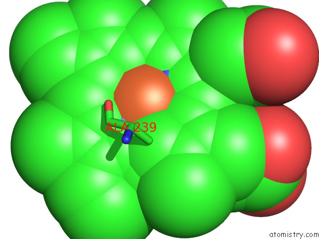
Mono view
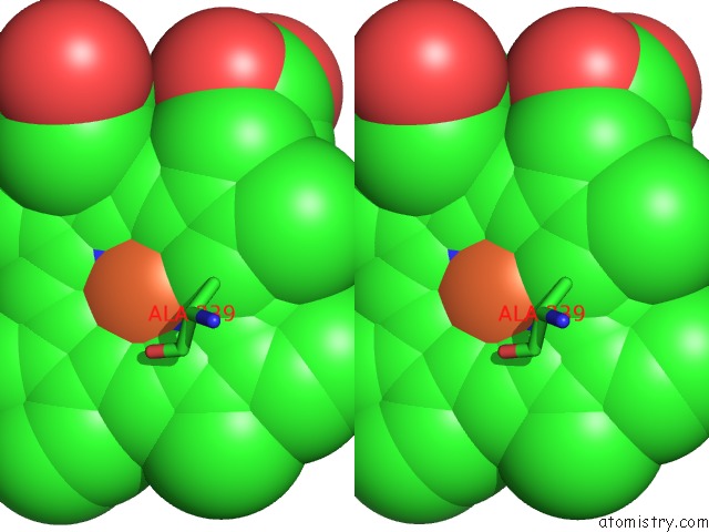
Stereo pair view
Mono view
Stereo pair view
A full contact list of Iron with other atoms in the Fe binding
site number 1 of Crystal Structure of Cytochrome P450 CYP107W1 From Streptomyces Avermitilis in Complex with Oligomycin A within 5.0Å range:
|
Reference:
L.W.Kang,
D.H.Kim,
T.V.Pham,
S.H.Han,
J.H.Kim,
Y.R.Lim,
H.G.Park,
G.S.Cha,
C.H.Yun,
Y.J.Chun.
Crystal Structure of Cytochrome P450 CYP107W1 From Streptomyces Avermitilis in Complex with Oligomycin A To Be Published.
Page generated: Tue Aug 5 16:50:28 2025
Last articles
Fe in 6GURFe in 6GPN
Fe in 6GM3
Fe in 6GM4
Fe in 6GPE
Fe in 6GMF
Fe in 6GM2
Fe in 6GM7
Fe in 6GM6
Fe in 6GM5