Iron »
PDB 4zoh-5ade »
4zxc »
Iron in PDB 4zxc: Crystal Structure of Hydroquinone 1,2-Dioxygenase Pnpcd in Complex with FE3+
Protein crystallography data
The structure of Crystal Structure of Hydroquinone 1,2-Dioxygenase Pnpcd in Complex with FE3+, PDB code: 4zxc
was solved by
S.Liu,
T.Su,
C.Zhang,
L.Gu,
with X-Ray Crystallography technique. A brief refinement statistics is given in the table below:
| Resolution Low / High (Å) | 33.12 / 3.05 |
| Space group | P 21 21 21 |
| Cell size a, b, c (Å), α, β, γ (°) | 76.837, 181.743, 186.863, 90.00, 90.00, 90.00 |
| R / Rfree (%) | 18.8 / 24.5 |
Iron Binding Sites:
The binding sites of Iron atom in the Crystal Structure of Hydroquinone 1,2-Dioxygenase Pnpcd in Complex with FE3+
(pdb code 4zxc). This binding sites where shown within
5.0 Angstroms radius around Iron atom.
In total 2 binding sites of Iron where determined in the Crystal Structure of Hydroquinone 1,2-Dioxygenase Pnpcd in Complex with FE3+, PDB code: 4zxc:
Jump to Iron binding site number: 1; 2;
In total 2 binding sites of Iron where determined in the Crystal Structure of Hydroquinone 1,2-Dioxygenase Pnpcd in Complex with FE3+, PDB code: 4zxc:
Jump to Iron binding site number: 1; 2;
Iron binding site 1 out of 2 in 4zxc
Go back to
Iron binding site 1 out
of 2 in the Crystal Structure of Hydroquinone 1,2-Dioxygenase Pnpcd in Complex with FE3+
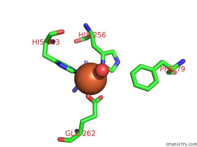
Mono view
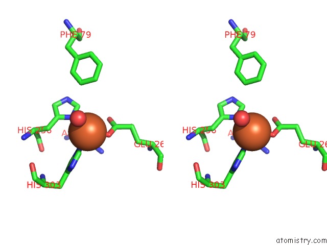
Stereo pair view
Mono view
Stereo pair view
A full contact list of Iron with other atoms in the Fe binding
site number 1 of Crystal Structure of Hydroquinone 1,2-Dioxygenase Pnpcd in Complex with FE3+ within 5.0Å range:
|
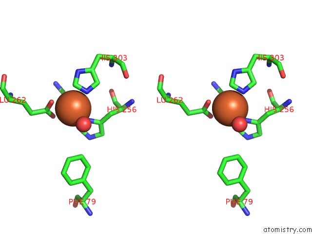
Iron binding site 2 out of 2 in 4zxc
Go back to
Iron binding site 2 out
of 2 in the Crystal Structure of Hydroquinone 1,2-Dioxygenase Pnpcd in Complex with FE3+
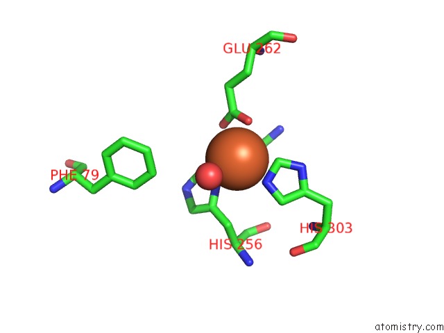
Mono view
Stereo pair view
Mono view
Stereo pair view
A full contact list of Iron with other atoms in the Fe binding
site number 2 of Crystal Structure of Hydroquinone 1,2-Dioxygenase Pnpcd in Complex with FE3+ within 5.0Å range:
|
Reference:
S.Liu,
T.Su,
C.Zhang,
W.M.Zhang,
D.Zhu,
J.Su,
T.Wei,
K.Wang,
Y.Huang,
L.Guo,
S.Xu,
N.Y.Zhou,
L.Gu.
Crystal Structure of Pnpcd, A Two-Subunit Hydroquinone 1,2-Dioxygenase, Reveals A Novel Structural Class of FE2+-Dependent Dioxygenases. J.Biol.Chem. V. 290 24547 2015.
ISSN: ESSN 1083-351X
PubMed: 26304122
DOI: 10.1074/JBC.M115.673558
Page generated: Tue Aug 5 18:39:47 2025
ISSN: ESSN 1083-351X
PubMed: 26304122
DOI: 10.1074/JBC.M115.673558
Last articles
Fe in 6LVVFe in 6LU1
Fe in 6LY4
Fe in 6LVC
Fe in 6LVB
Fe in 6LS3
Fe in 6LS8
Fe in 6LTM
Fe in 6LTL
Fe in 6LS0