Iron »
PDB 5why-5xbp »
5wjk »
Iron in PDB 5wjk: 2.0-Angstrom in Situ Mylar Structure of Sperm Whale Myoglobin (Swmb) at 293 K
Protein crystallography data
The structure of 2.0-Angstrom in Situ Mylar Structure of Sperm Whale Myoglobin (Swmb) at 293 K, PDB code: 5wjk
was solved by
J.Broecker,
W.-L.Ou,
O.P.Ernst,
with X-Ray Crystallography technique. A brief refinement statistics is given in the table below:
| Resolution Low / High (Å) | 45.84 / 2.00 |
| Space group | P 6 |
| Cell size a, b, c (Å), α, β, γ (°) | 91.670, 91.670, 46.170, 90.00, 90.00, 120.00 |
| R / Rfree (%) | 22.4 / 26.9 |
Other elements in 5wjk:
The structure of 2.0-Angstrom in Situ Mylar Structure of Sperm Whale Myoglobin (Swmb) at 293 K also contains other interesting chemical elements:
| Chlorine | (Cl) | 5 atoms |
Iron Binding Sites:
The binding sites of Iron atom in the 2.0-Angstrom in Situ Mylar Structure of Sperm Whale Myoglobin (Swmb) at 293 K
(pdb code 5wjk). This binding sites where shown within
5.0 Angstroms radius around Iron atom.
In total only one binding site of Iron was determined in the 2.0-Angstrom in Situ Mylar Structure of Sperm Whale Myoglobin (Swmb) at 293 K, PDB code: 5wjk:
In total only one binding site of Iron was determined in the 2.0-Angstrom in Situ Mylar Structure of Sperm Whale Myoglobin (Swmb) at 293 K, PDB code: 5wjk:
Iron binding site 1 out of 1 in 5wjk
Go back to
Iron binding site 1 out
of 1 in the 2.0-Angstrom in Situ Mylar Structure of Sperm Whale Myoglobin (Swmb) at 293 K
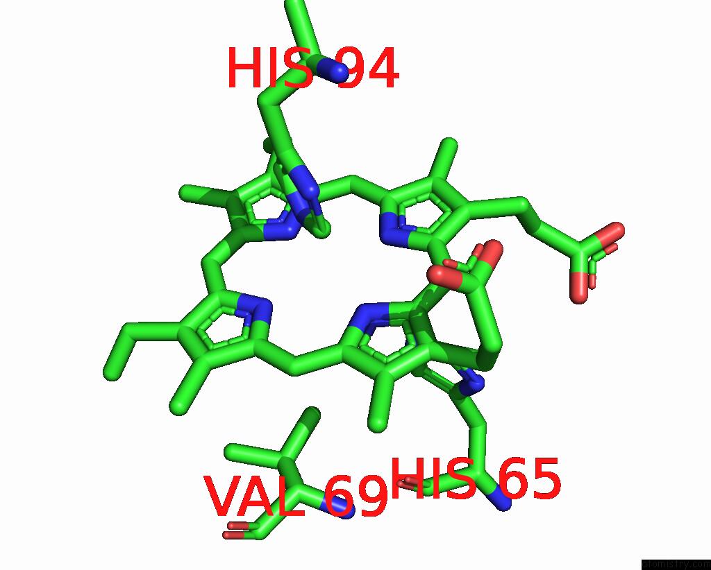
Mono view
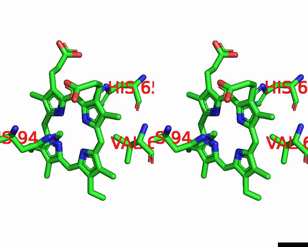
Stereo pair view
Mono view
Stereo pair view
A full contact list of Iron with other atoms in the Fe binding
site number 1 of 2.0-Angstrom in Situ Mylar Structure of Sperm Whale Myoglobin (Swmb) at 293 K within 5.0Å range:
|
Reference:
J.Broecker,
T.Morizumi,
W.L.Ou,
V.Klingel,
A.Kuo,
D.J.Kissick,
A.Ishchenko,
M.Y.Lee,
S.Xu,
O.Makarov,
V.Cherezov,
C.M.Ogata,
O.P.Ernst.
High-Throughput in Situ X-Ray Screening of and Data Collection From Protein Crystals at Room Temperature and Under Cryogenic Conditions. Nat Protoc V. 13 260 2018.
ISSN: ESSN 1750-2799
PubMed: 29300389
DOI: 10.1038/NPROT.2017.135
Page generated: Wed Aug 6 02:31:33 2025
ISSN: ESSN 1750-2799
PubMed: 29300389
DOI: 10.1038/NPROT.2017.135
Last articles
Fe in 6CXUFe in 6CSB
Fe in 6CWW
Fe in 6CVC
Fe in 6CUN
Fe in 6CTC
Fe in 6CSD
Fe in 6CUK
Fe in 6CIP
Fe in 6CIN