Iron »
PDB 5xzi-5ylw »
5y5m »
Iron in PDB 5y5m: Sfx Structure of Cytochrome P450NOR: A Complete Dark Data Without Pump Laser (Resting State)
Enzymatic activity of Sfx Structure of Cytochrome P450NOR: A Complete Dark Data Without Pump Laser (Resting State)
All present enzymatic activity of Sfx Structure of Cytochrome P450NOR: A Complete Dark Data Without Pump Laser (Resting State):
1.7.1.14;
1.7.1.14;
Protein crystallography data
The structure of Sfx Structure of Cytochrome P450NOR: A Complete Dark Data Without Pump Laser (Resting State), PDB code: 5y5m
was solved by
T.Tosha,
T.Nomura,
T.Nishida,
N.Saeki,
K.Okubayashi,
R.Yamagiwa,
M.Sugahara,
T.Nakane,
K.Yamashita,
K.Hirata,
G.Ueno,
T.Kimura,
T.Hisano,
K.Muramoto,
H.Sawai,
H.Takeda,
E.Mizohata,
A.Yamashita,
Y.Kanematsu,
Y.Takano,
E.Nango,
R.Tanaka,
O.Nureki,
Y.Ikemoto,
H.Murakami,
S.Owada,
K.Tono,
M.Yabashi,
M.Yamamoto,
H.Ago,
S.Iwata,
H.Sugimoto,
Y.Shiro,
M.Kubo,
with X-Ray Crystallography technique. A brief refinement statistics is given in the table below:
| Resolution Low / High (Å) | 19.92 / 2.10 |
| Space group | P 1 21 1 |
| Cell size a, b, c (Å), α, β, γ (°) | 54.600, 102.300, 73.700, 90.00, 92.60, 90.00 |
| R / Rfree (%) | 16.8 / 21.8 |
Iron Binding Sites:
The binding sites of Iron atom in the Sfx Structure of Cytochrome P450NOR: A Complete Dark Data Without Pump Laser (Resting State)
(pdb code 5y5m). This binding sites where shown within
5.0 Angstroms radius around Iron atom.
In total 2 binding sites of Iron where determined in the Sfx Structure of Cytochrome P450NOR: A Complete Dark Data Without Pump Laser (Resting State), PDB code: 5y5m:
Jump to Iron binding site number: 1; 2;
In total 2 binding sites of Iron where determined in the Sfx Structure of Cytochrome P450NOR: A Complete Dark Data Without Pump Laser (Resting State), PDB code: 5y5m:
Jump to Iron binding site number: 1; 2;
Iron binding site 1 out of 2 in 5y5m
Go back to
Iron binding site 1 out
of 2 in the Sfx Structure of Cytochrome P450NOR: A Complete Dark Data Without Pump Laser (Resting State)
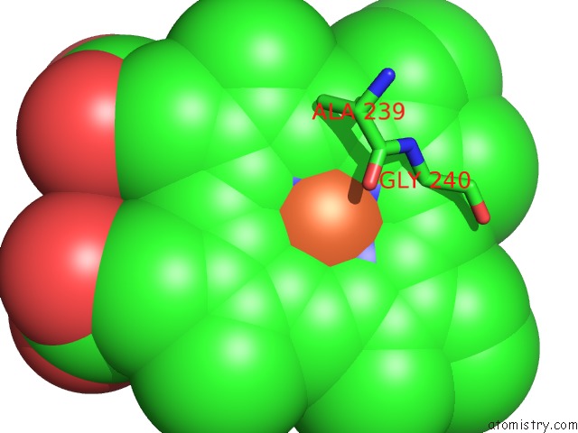
Mono view
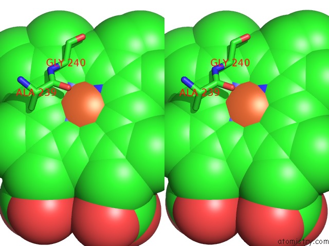
Stereo pair view
Mono view
Stereo pair view
A full contact list of Iron with other atoms in the Fe binding
site number 1 of Sfx Structure of Cytochrome P450NOR: A Complete Dark Data Without Pump Laser (Resting State) within 5.0Å range:
|
Iron binding site 2 out of 2 in 5y5m
Go back to
Iron binding site 2 out
of 2 in the Sfx Structure of Cytochrome P450NOR: A Complete Dark Data Without Pump Laser (Resting State)
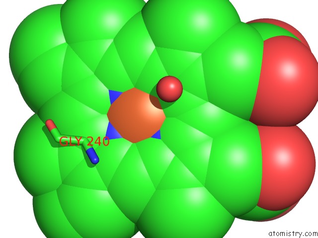
Mono view
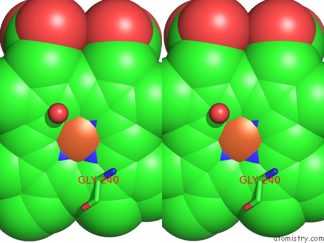
Stereo pair view
Mono view
Stereo pair view
A full contact list of Iron with other atoms in the Fe binding
site number 2 of Sfx Structure of Cytochrome P450NOR: A Complete Dark Data Without Pump Laser (Resting State) within 5.0Å range:
|
Reference:
T.Tosha,
T.Nomura,
T.Nishida,
N.Saeki,
K.Okubayashi,
R.Yamagiwa,
M.Sugahara,
T.Nakane,
K.Yamashita,
K.Hirata,
G.Ueno,
T.Kimura,
T.Hisano,
K.Muramoto,
H.Sawai,
H.Takeda,
E.Mizohata,
A.Yamashita,
Y.Kanematsu,
Y.Takano,
E.Nango,
R.Tanaka,
O.Nureki,
O.Shoji,
Y.Ikemoto,
H.Murakami,
S.Owada,
K.Tono,
M.Yabashi,
M.Yamamoto,
H.Ago,
S.Iwata,
H.Sugimoto,
Y.Shiro,
M.Kubo.
Capturing An Initial Intermediate During the P450NOR Enzymatic Reaction Using Time-Resolved Xfel Crystallography and Caged-Substrate. Nat Commun V. 8 1585 2017.
ISSN: ESSN 2041-1723
PubMed: 29147002
DOI: 10.1038/S41467-017-01702-1
Page generated: Wed Aug 6 03:20:16 2025
ISSN: ESSN 2041-1723
PubMed: 29147002
DOI: 10.1038/S41467-017-01702-1
Last articles
Fe in 7AQQFe in 7AO7
Fe in 7ANV
Fe in 7ANT
Fe in 7AKT
Fe in 7AI9
Fe in 7AIK
Fe in 7AIL
Fe in 7ADY
Fe in 7ADR