Iron »
PDB 7dyb-7ek5 »
7ef9 »
Iron in PDB 7ef9: Crystal Structure of Mouse Mutyh in Complex with Dna Containing Ap Site Analogue:8-Oxog (Form II)
Enzymatic activity of Crystal Structure of Mouse Mutyh in Complex with Dna Containing Ap Site Analogue:8-Oxog (Form II)
All present enzymatic activity of Crystal Structure of Mouse Mutyh in Complex with Dna Containing Ap Site Analogue:8-Oxog (Form II):
3.2.2.31;
3.2.2.31;
Protein crystallography data
The structure of Crystal Structure of Mouse Mutyh in Complex with Dna Containing Ap Site Analogue:8-Oxog (Form II), PDB code: 7ef9
was solved by
T.Nakamura,
Y.Nakabeppu,
Y.Yamagata,
with X-Ray Crystallography technique. A brief refinement statistics is given in the table below:
| Resolution Low / High (Å) | 42.53 / 1.97 |
| Space group | I 2 2 2 |
| Cell size a, b, c (Å), α, β, γ (°) | 71.66, 108.496, 158.527, 90, 90, 90 |
| R / Rfree (%) | 17.8 / 19.7 |
Other elements in 7ef9:
The structure of Crystal Structure of Mouse Mutyh in Complex with Dna Containing Ap Site Analogue:8-Oxog (Form II) also contains other interesting chemical elements:
| Zinc | (Zn) | 1 atom |
Iron Binding Sites:
The binding sites of Iron atom in the Crystal Structure of Mouse Mutyh in Complex with Dna Containing Ap Site Analogue:8-Oxog (Form II)
(pdb code 7ef9). This binding sites where shown within
5.0 Angstroms radius around Iron atom.
In total 4 binding sites of Iron where determined in the Crystal Structure of Mouse Mutyh in Complex with Dna Containing Ap Site Analogue:8-Oxog (Form II), PDB code: 7ef9:
Jump to Iron binding site number: 1; 2; 3; 4;
In total 4 binding sites of Iron where determined in the Crystal Structure of Mouse Mutyh in Complex with Dna Containing Ap Site Analogue:8-Oxog (Form II), PDB code: 7ef9:
Jump to Iron binding site number: 1; 2; 3; 4;
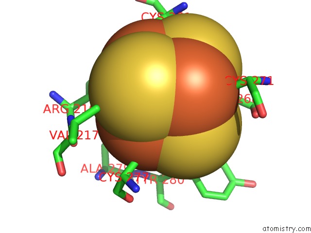
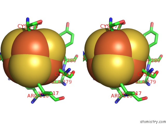
Iron binding site 1 out of 4 in 7ef9
Go back to
Iron binding site 1 out
of 4 in the Crystal Structure of Mouse Mutyh in Complex with Dna Containing Ap Site Analogue:8-Oxog (Form II)
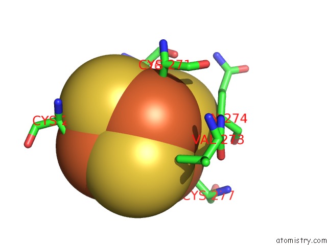
Mono view
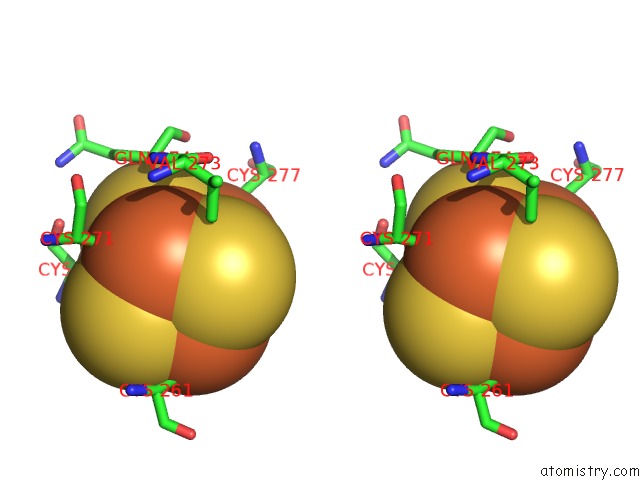
Stereo pair view
Mono view
Stereo pair view
A full contact list of Iron with other atoms in the Fe binding
site number 1 of Crystal Structure of Mouse Mutyh in Complex with Dna Containing Ap Site Analogue:8-Oxog (Form II) within 5.0Å range:
|
Iron binding site 2 out of 4 in 7ef9
Go back to
Iron binding site 2 out
of 4 in the Crystal Structure of Mouse Mutyh in Complex with Dna Containing Ap Site Analogue:8-Oxog (Form II)
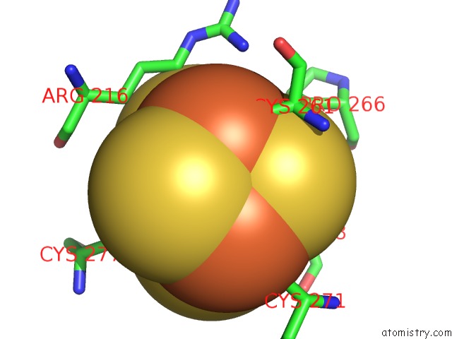
Mono view
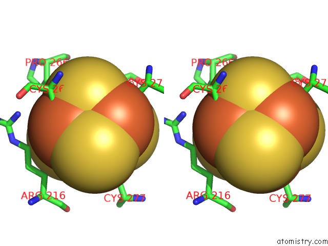
Stereo pair view
Mono view
Stereo pair view
A full contact list of Iron with other atoms in the Fe binding
site number 2 of Crystal Structure of Mouse Mutyh in Complex with Dna Containing Ap Site Analogue:8-Oxog (Form II) within 5.0Å range:
|
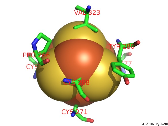
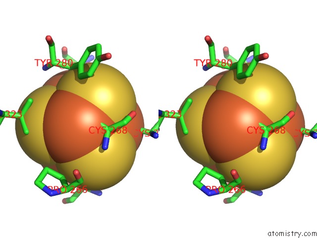
Iron binding site 3 out of 4 in 7ef9
Go back to
Iron binding site 3 out
of 4 in the Crystal Structure of Mouse Mutyh in Complex with Dna Containing Ap Site Analogue:8-Oxog (Form II)
Mono view
Stereo pair view
Mono view
Stereo pair view
A full contact list of Iron with other atoms in the Fe binding
site number 3 of Crystal Structure of Mouse Mutyh in Complex with Dna Containing Ap Site Analogue:8-Oxog (Form II) within 5.0Å range:
|
Iron binding site 4 out of 4 in 7ef9
Go back to
Iron binding site 4 out
of 4 in the Crystal Structure of Mouse Mutyh in Complex with Dna Containing Ap Site Analogue:8-Oxog (Form II)
Mono view
Stereo pair view
Mono view
Stereo pair view
A full contact list of Iron with other atoms in the Fe binding
site number 4 of Crystal Structure of Mouse Mutyh in Complex with Dna Containing Ap Site Analogue:8-Oxog (Form II) within 5.0Å range:
|
Reference:
T.Nakamura,
K.Okabe,
S.Hirayama,
M.Chirifu,
S.Ikemizu,
H.Morioka,
Y.Nakabeppu,
Y.Yamagata.
Structure of the Mammalian Adenine Dna Glycosylase Mutyh: Insights Into the Base Excision Repair Pathway and Cancer. Nucleic Acids Res. 2021.
ISSN: ESSN 1362-4962
DOI: 10.1093/NAR/GKAB492
Page generated: Wed Aug 6 22:13:39 2025
ISSN: ESSN 1362-4962
DOI: 10.1093/NAR/GKAB492
Last articles
Fe in 8DVYFe in 8DVP
Fe in 8DPN
Fe in 8DQV
Fe in 8DVX
Fe in 8DSG
Fe in 8DNP
Fe in 8DRM
Fe in 8DRL
Fe in 8DRJ