Iron »
PDB 7fcb-7k5g »
7jj1 »
Iron in PDB 7jj1: Crystal Structure of the Sterol 14ALPHA-Demethylase-Ferredoxin (CYP51- Fx) Heme Domain and Architectural Comparison to the Whole Fusion Protein
Enzymatic activity of Crystal Structure of the Sterol 14ALPHA-Demethylase-Ferredoxin (CYP51- Fx) Heme Domain and Architectural Comparison to the Whole Fusion Protein
All present enzymatic activity of Crystal Structure of the Sterol 14ALPHA-Demethylase-Ferredoxin (CYP51- Fx) Heme Domain and Architectural Comparison to the Whole Fusion Protein:
1.14.13.70;
1.14.13.70;
Protein crystallography data
The structure of Crystal Structure of the Sterol 14ALPHA-Demethylase-Ferredoxin (CYP51- Fx) Heme Domain and Architectural Comparison to the Whole Fusion Protein, PDB code: 7jj1
was solved by
B.Zhao,
D.C.Lamb,
with X-Ray Crystallography technique. A brief refinement statistics is given in the table below:
| Resolution Low / High (Å) | 29.52 / 3.00 |
| Space group | C 2 2 21 |
| Cell size a, b, c (Å), α, β, γ (°) | 157.682, 164.755, 187.683, 90, 90, 90 |
| R / Rfree (%) | 19.6 / 23.7 |
Iron Binding Sites:
The binding sites of Iron atom in the Crystal Structure of the Sterol 14ALPHA-Demethylase-Ferredoxin (CYP51- Fx) Heme Domain and Architectural Comparison to the Whole Fusion Protein
(pdb code 7jj1). This binding sites where shown within
5.0 Angstroms radius around Iron atom.
In total 3 binding sites of Iron where determined in the Crystal Structure of the Sterol 14ALPHA-Demethylase-Ferredoxin (CYP51- Fx) Heme Domain and Architectural Comparison to the Whole Fusion Protein, PDB code: 7jj1:
Jump to Iron binding site number: 1; 2; 3;
In total 3 binding sites of Iron where determined in the Crystal Structure of the Sterol 14ALPHA-Demethylase-Ferredoxin (CYP51- Fx) Heme Domain and Architectural Comparison to the Whole Fusion Protein, PDB code: 7jj1:
Jump to Iron binding site number: 1; 2; 3;
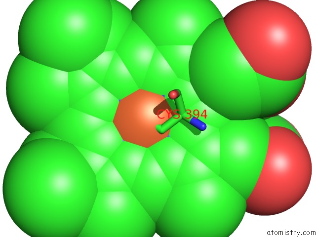
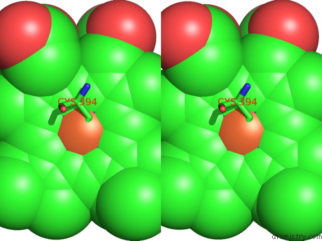
Iron binding site 1 out of 3 in 7jj1
Go back to
Iron binding site 1 out
of 3 in the Crystal Structure of the Sterol 14ALPHA-Demethylase-Ferredoxin (CYP51- Fx) Heme Domain and Architectural Comparison to the Whole Fusion Protein
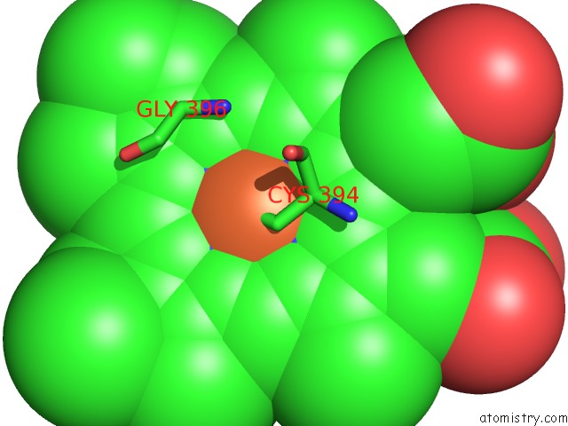
Mono view
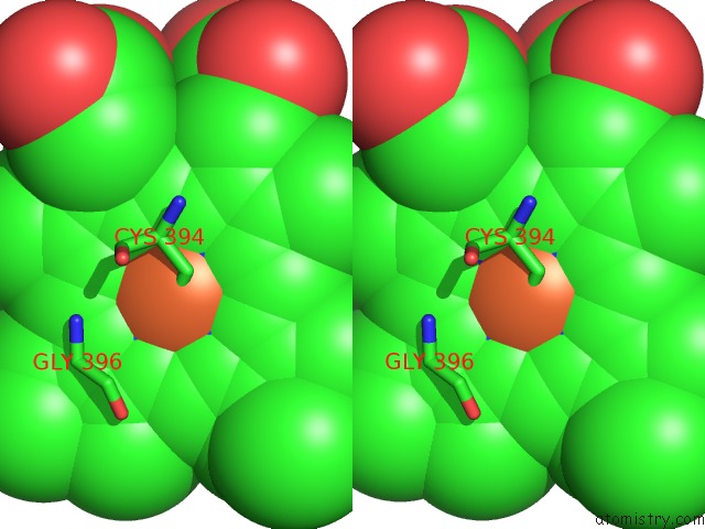
Stereo pair view
Mono view
Stereo pair view
A full contact list of Iron with other atoms in the Fe binding
site number 1 of Crystal Structure of the Sterol 14ALPHA-Demethylase-Ferredoxin (CYP51- Fx) Heme Domain and Architectural Comparison to the Whole Fusion Protein within 5.0Å range:
|
Iron binding site 2 out of 3 in 7jj1
Go back to
Iron binding site 2 out
of 3 in the Crystal Structure of the Sterol 14ALPHA-Demethylase-Ferredoxin (CYP51- Fx) Heme Domain and Architectural Comparison to the Whole Fusion Protein
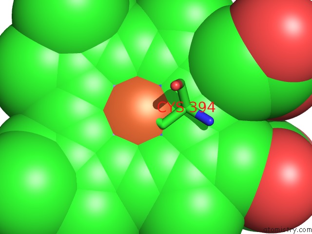
Mono view
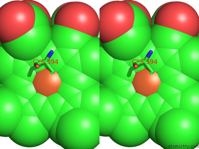
Stereo pair view
Mono view
Stereo pair view
A full contact list of Iron with other atoms in the Fe binding
site number 2 of Crystal Structure of the Sterol 14ALPHA-Demethylase-Ferredoxin (CYP51- Fx) Heme Domain and Architectural Comparison to the Whole Fusion Protein within 5.0Å range:
|
Iron binding site 3 out of 3 in 7jj1
Go back to
Iron binding site 3 out
of 3 in the Crystal Structure of the Sterol 14ALPHA-Demethylase-Ferredoxin (CYP51- Fx) Heme Domain and Architectural Comparison to the Whole Fusion Protein
Mono view
Stereo pair view
Mono view
Stereo pair view
A full contact list of Iron with other atoms in the Fe binding
site number 3 of Crystal Structure of the Sterol 14ALPHA-Demethylase-Ferredoxin (CYP51- Fx) Heme Domain and Architectural Comparison to the Whole Fusion Protein within 5.0Å range:
|
Reference:
B.Zhao,
D.C.Lamb.
Crystal Structure of the Sterol 14ALPHA-Demethylase-Ferredoxin (CYP51-Fx) Heme Domain and Architectural Comparison to the Whole Fusion Protein To Be Published.
Page generated: Wed Aug 6 22:28:24 2025
Last articles
Fe in 8DW4Fe in 8DVY
Fe in 8DVP
Fe in 8DPN
Fe in 8DQV
Fe in 8DVX
Fe in 8DSG
Fe in 8DNP
Fe in 8DRM
Fe in 8DRL